. 2011 Jul;21(7):1526-34.
doi: 10.1007/s00330-011-2081-y. Epub 2011 Feb 18.

Early postoperative MRI overestimates residual tumour after resection of gliomas with no or minimal enhancement

Affiliations

Early postoperative MRI overestimates residual tumour after resection of gliomas with no or minimal enhancement

Sinan M K Belhawi et al. Eur Radiol. 2011 Jul.

Abstract

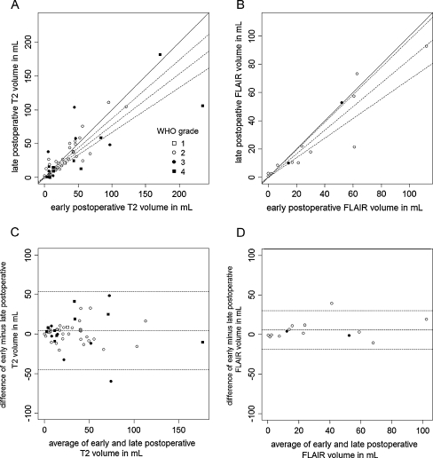
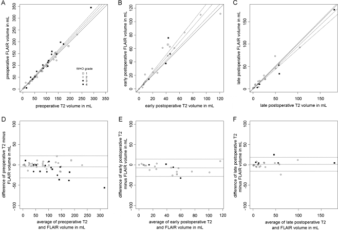
Background: Standards for residual tumour measurement after resection of gliomas with no or minimal enhancement have not yet been established. In this study residual volumes on early and late postoperative T2-/FLAIR-weighted MRI are compared.

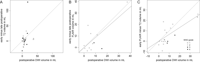
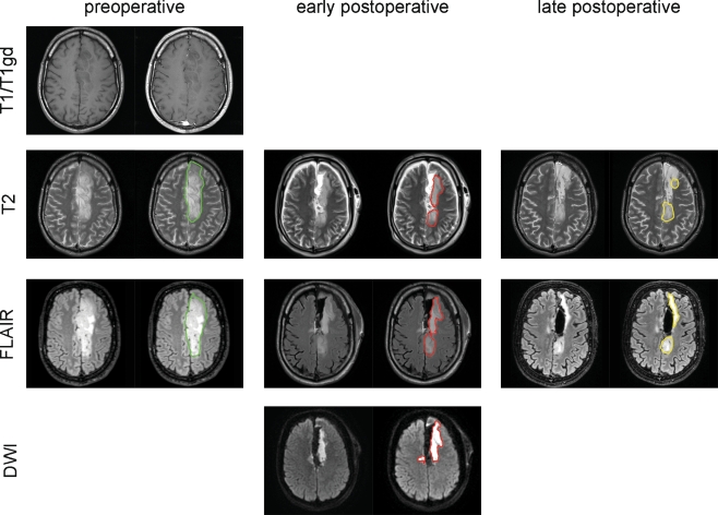
Methods: A retrospective cohort included 58 consecutive glioma patients with no or minimal preoperative gadolinium enhancement. Inclusion criteria were first-time resection between 2007 and 2009 with a T2-/FLAIR-based target volume and availability of preoperative, early (<48 h) and late (1-7 months) postoperative MRI. The volumes of non-enhancing T2/FLAIR tissue and diffusion restriction areas were measured.

Results: Residual tumour volumes were 22% smaller on late postoperative compared with early postoperative T2-weighted MRI and 49% smaller for FLAIR-weighted imaging. Postoperative restricted diffusion volume correlated with the difference between early and late postoperative FLAIR volumes and with the difference between T2 and FLAIR volumes on early postoperative MRI.

Conclusion: We observed a systematic and substantial overestimation of residual non-enhancing volume on MRI within 48 h of resection compared with months postoperatively, in particular for FLAIR imaging. Resection-induced ischaemia contributes to this overestimation, as may other operative effects. This indicates that early postoperative MRI is less reliable to determine the extent of non-enhancing residual glioma and restricted diffusion volumes are imperative.


Figures

Fig. 1
Fig. 1
Residual tumour volumes in mL comparing early and late postoperative MRI based on T2-weighted imaging and FLAIR-weighted imaging shown as data plots (a and b) and Bland-Altman plots (c and d) respectively depicting systematic overestimation of residual tumour volume by early postoperative MRI. Each data point represents measurements obtained from one patient, tagged by glioma grade according to the legend. The straight diagonal line in a and b represents hypothetical perfect agreement and the dotted lines the actual linear regression fit and corresponding 95% confidence interval. The three dotted horizontal lines in c and d represent the average differential volume and corresponding 95% confidence interval
Fig. 2
Fig. 2
Residual tumour volumes in mL comparing T2- and FLAIR-weighted imaging respectively based on preoperative, early postoperative and late postoperative MRI shown as data plots (a, b and c) and Bland Altman plots (d, e and f) depicting good agreement in residual tumour volume based on preoperative and late postoperative MRI and systematic overestimation of FLAIR-weighted imaging on early postoperative MRI. Data points, tagging and line styles as in Fig. 1
Fig. 3
Fig. 3
Plots of diffusion restriction volume on early postoperative MRI and (a) the difference in T2 residual tumour volumes of early and late postoperative MRI, (b) the difference in FLAIR residual tumour volumes of early and late postoperative MRI and (c) the difference in early postoperative MRI residual tumour volumes of T2- and FLAIR-weighted imaging. Data points, tagging and line styles as in Fig. 1
Fig. 4
Fig. 4
An example of preoperative, early and late postoperative MRI displaying T2-, FLAIR- and diffusion-weighted axial images and volume contours. This 36-year old man with an oligoastrocytoma in the left cingulate gyrus had preoperative T2 and FLAIR tumour volumes of 101 and 94 cm3, respectively, outlined in green, with faint (2%) T1 gadolinium enhancement 3 weeks before resection. The residual T2 and FLAIR volumes 2 days after surgery were 35 and 61 cm3, respectively, outlined in red, with a diffusion restriction volume of 37 cm3. The residual T2 and FLAIR volumes 97 days after resection measured 27 and 21 cm3, respectively, outlined in yellow. Note the involution of the hyperintensity at the lateral margin of the resection cavity with restricted diffusion, interpreted as resection-induced ischaemia, and the stable hyperintensity at the posterior margin of the resection cavity, presumably genuine residual glioma within the corticospinal tract

References

    1. Gorlia T, van den Bent MJ, Hegi ME, et al. Nomograms for predicting survival of patients with newly diagnosed glioblastoma: prognostic factor analysis of EORTC and NCIC trial 26981-22981/CE.3. Lancet Oncol. 2008;9:29–38. doi: 10.1016/S1470-2045(07)70384-4. - DOI - PubMed
    1. Chang EF, Smith JS, Chang SM, et al. Preoperative prognostic classification system for hemispheric low-grade gliomas in adults. J Neurosurg. 2008;109:817–824. doi: 10.3171/JNS/2008/109/11/0817. - DOI - PubMed
    1. McGirt MJ, Chaichana KL, Attenello FJ, et al. Extent of surgical resection is independently associated with survival in patients with hemispheric infiltrating low-grade gliomas. Neurosurgery. 2008;63:700–707. doi: 10.1227/01.NEU.0000325729.41085.73. - DOI - PubMed
    1. Smith JS, Chang EF, Lamborn KR, et al. Role of extent of resection in the long-term outcome of low-grade hemispheric gliomas. J Clin Oncol. 2008;26:1338–1345. doi: 10.1200/JCO.2007.13.9337. - DOI - PubMed
    1. Stummer W, Reulen HJ, Meinel T, et al. Extent of resection and survival in glioblastoma multiforme: identification of and adjustment for bias. Neurosurgery. 2008;62:564–576. doi: 10.1227/01.neu.0000317304.31579.17. - DOI - PubMed