

Spatial frequency domain imaging of intrinsic optical property contrast in a mouse model of Alzheimer's disease
- PMID: 21331663
- PMCID: PMC3069335
- DOI: 10.1007/s10439-011-0269-6
Spatial frequency domain imaging of intrinsic optical property contrast in a mouse model of Alzheimer's disease
Abstract
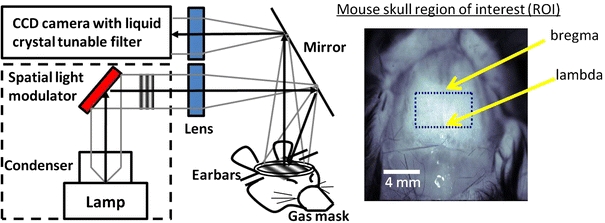
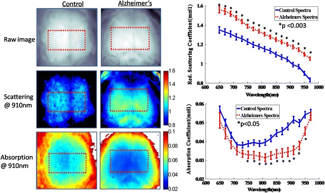
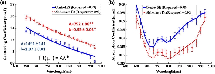
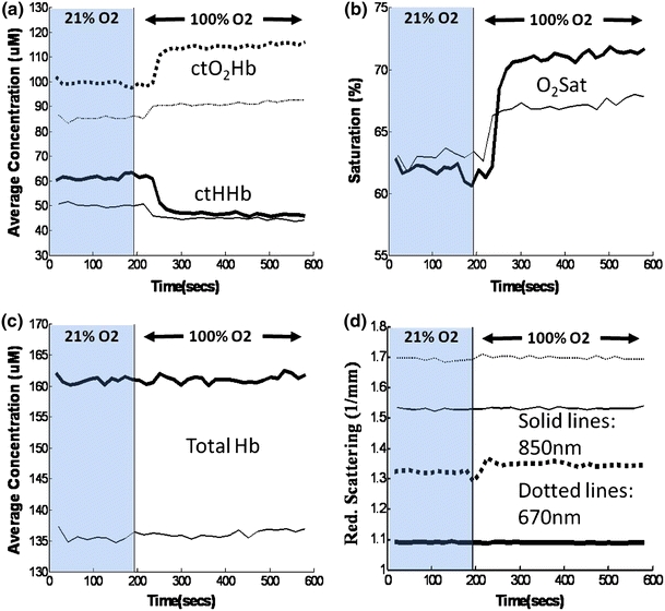
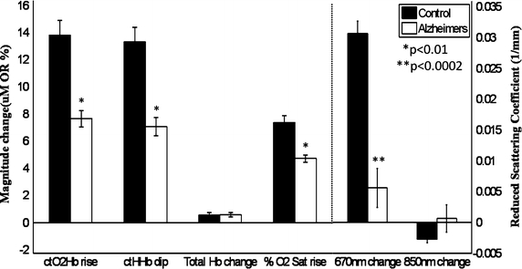
Extensive changes in neural tissue structure and function accompanying Alzheimer's disease (AD) suggest that intrinsic signal optical imaging can provide new contrast mechanisms and insight for assessing AD appearance and progression. In this work, we report the development of a wide-field spatial frequency domain imaging (SFDI) method for non-contact, quantitative in vivo optical imaging of brain tissue composition and function in a triple transgenic mouse AD model (3xTg). SFDI was used to generate optical absorption and scattering maps at up to 17 wavelengths from 650 to 970 nm in 20-month-old 3xTg mice (n = 4) and age-matched controls (n = 6). Wavelength-dependent optical properties were used to form images of tissue hemoglobin (oxy-, deoxy-, and total), oxygen saturation, and water. Significant baseline contrast was observed with 13-26% higher average scattering values and elevated water content (52 ± 2% vs. 31 ± 1%); reduced total tissue hemoglobin content (127 ± 9 μM vs. 174 ± 6 μM); and lower tissue oxygen saturation (57 ± 2% vs. 69 ± 3%) in AD vs. control mice. Oxygen inhalation challenges (100% oxygen) resulted in increased levels of tissue oxy-hemoglobin (ctO(2)Hb) and commensurate reductions in deoxy-hemoglobin (ctHHb), with ~60-70% slower response times and ~7 μM vs. ~14 μM overall changes for 3xTg vs. controls, respectively. Our results show that SFDI is capable of revealing quantitative functional contrast in an AD model and may be a useful method for studying dynamic alterations in AD neural tissue composition and physiology.
Figures
Similar articles
-
Characterization of a 3xTg-AD mouse model of Alzheimer's disease with the senescence accelerated mouse prone 8 (SAMP8) background.Synapse. 2018 Apr;72(4). doi: 10.1002/syn.22025. Epub 2018 Feb 1. Synapse. 2018. PMID: 29341269
-
Spatial frequency domain imaging of port wine stain biochemical composition in response to laser therapy: a pilot study.Lasers Surg Med. 2012 Oct;44(8):611-21. doi: 10.1002/lsm.22067. Epub 2012 Aug 21. Lasers Surg Med. 2012. PMID: 22911574 Free PMC article.
-
Selenomethionine Mitigates Cognitive Decline by Targeting Both Tau Hyperphosphorylation and Autophagic Clearance in an Alzheimer's Disease Mouse Model.J Neurosci. 2017 Mar 1;37(9):2449-2462. doi: 10.1523/JNEUROSCI.3229-16.2017. Epub 2017 Jan 30. J Neurosci. 2017. PMID: 28137967 Free PMC article.
-
Alcohol drinking exacerbates neural and behavioral pathology in the 3xTg-AD mouse model of Alzheimer's disease.Int Rev Neurobiol. 2019;148:169-230. doi: 10.1016/bs.irn.2019.10.017. Epub 2019 Oct 23. Int Rev Neurobiol. 2019. PMID: 31733664 Free PMC article. Review.
-
Quantitative In Vivo Imaging of Tissue Absorption, Scattering, and Hemoglobin Concentration in Rat Cortex Using Spatially Modulated Structured Light.In: Frostig RD, editor. In Vivo Optical Imaging of Brain Function. 2nd edition. Boca Raton (FL): CRC Press/Taylor & Francis; 2009. Chapter 12. In: Frostig RD, editor. In Vivo Optical Imaging of Brain Function. 2nd edition. Boca Raton (FL): CRC Press/Taylor & Francis; 2009. Chapter 12. PMID: 26844326 Free Books & Documents. Review.
Cited by
-
Shortwave-infrared meso-patterned imaging enables label-free mapping of tissue water and lipid content.Nat Commun. 2020 Oct 23;11(1):5355. doi: 10.1038/s41467-020-19128-7. Nat Commun. 2020. PMID: 33097705 Free PMC article.
-
Deep learning in macroscopic diffuse optical imaging.J Biomed Opt. 2022 Feb;27(2):020901. doi: 10.1117/1.JBO.27.2.020901. J Biomed Opt. 2022. PMID: 35218169 Free PMC article. Review.
-
Microvascular imaging of the skin.Phys Med Biol. 2019 Mar 21;64(7):07TR01. doi: 10.1088/1361-6560/ab03f1. Phys Med Biol. 2019. PMID: 30708364 Free PMC article. Review.
-
Optical imaging in an Alzheimer's mouse model reveals amyloid-β-dependent vascular impairment.Neurophotonics. 2014 Jul;1(1):011005. doi: 10.1117/1.NPh.1.1.011005. Neurophotonics. 2014. PMID: 25133200 Free PMC article.
-
Small separation frequency-domain near-infrared spectroscopy for the recovery of tissue optical properties at millimeter depths.Biomed Opt Express. 2019 Sep 27;10(10):5362-5377. doi: 10.1364/BOE.10.005362. eCollection 2019 Oct 1. Biomed Opt Express. 2019. PMID: 31646051 Free PMC article.
References
Publication types
MeSH terms
Substances
Grants and funding
LinkOut - more resources
Full Text Sources
Other Literature Sources
Medical
