. 2011 Mar;31(3):289-93.
doi: 10.1007/s10059-011-0038-2. Epub 2011 Feb 10.

CaMKII regulates pericyte loss in the retina of early diabetic mouse

Affiliations

CaMKII regulates pericyte loss in the retina of early diabetic mouse

Young Hee Kim et al. Mol Cells. 2011 Mar.

Abstract

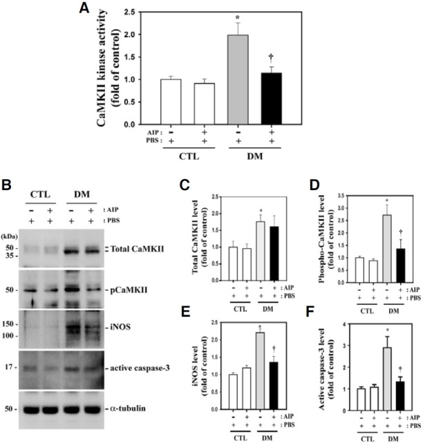
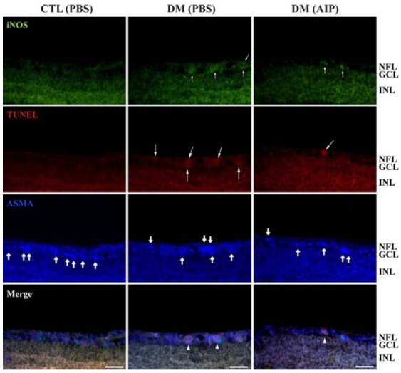
Inducible nitric oxide synthase (iNOS) is an essential mediator in diabetic vascular lesions and known to be regulated by activation of Ca(2+)/calmodulin-dependent protein kinase II (CaMKII). The aim of this study was to investigate whether CaMKII affects iNOS-mediated pericyte death in the retina of diabetic mice with early stage disease. Total- and phospho-CaMKII, iNOS, and active caspase-3 protein levels were assessed by Western blotting, and CaMKII activity was measured by kinase assay. iNOS-related pericyte death was assessed by double immunofluorescent staining for iNOS and α-smooth muscle actin, followed by the TUNEL assay. Autocamtide-2-related inhibitory peptide (AIP), a specific inhibitor of CaMKII, was injected into the right vitreous 2 days before sacrifice of mice, to examine the effect of CaMKII inactivation in diabetic retinas. The levels of total- and phospho-CaMKII, iNOS, and active caspase-3 protein, and CaMKII activity were significantly increased in the diabetic retinas compared with those of control retinas. Furthermore, TUNEL-positive signals colocalized with iNOS-immunoreactive pericytes in the same retinas. However, inactivation of CaMKII by AIP treatment inhibited all these changes, which was accompanied by less pericyte loss. Our results demonstrate that CaMKII contributes to iNOS-related death of pericytes in the diabetic retina and that inactivation of this enzyme may be a potential treatment for retinal vascular lesion.


Figures

Fig. 1.
Fig. 1.. CaMKII and caspase-3 activity and total CaMKII and iNOS levels in diabetic mouse retina and the effect of AIP treatment. (A) CaMKII kinase activity and the levels of these proteins increased in the retinas of diabetic mice compared with the controls (*P = 0.013, n = 4). Activity was greatly reduced in the retinas of AIPtreated diabetic mice compared with PBS-treated diabetic mice (P = 0.033, n = 4). (B) Western blot analysis using the indicated specific antibodies on total cell lysates of retinas from diabetic and control mice. Caspase- 3, iNOS and phospho-CaMKII levels were reduced in AIP-treated diabetic mice, whereas total CaMKII protein levels did not significantly change (P = 0.813, n = 4). (C-F) Quantitative representation of Western blot results for the indicated proteins. All experiments were performed in quadruplicate. Data are presented as the mean ± SEM (n = 4). *P < 0.05, comparing the PBS-treated control and diabetic mice; P < 0.05, comparing the PBS- and AIP-treated diabetic groups.
Fig. 2.
Fig. 2.. Correlation of CaMKII activity and iNOS expression with pericyte death in diabetic mice retinas. Double immunofluorescent staining for iNOS and ASMA (pericyte marker) was performed, followed by the TUNEL assay with TMR red on the same frozen sections. Increased iNOS and TUNEL-positive cells were observed in diabetic mice retinas (small and large arrows). These signals co-localized with ASMA-positive pericytes (thick arrows). The diabetes-induced increase in iNOS-positive dead pericytes (arrowheads) was suppressed by AIP treatment. GCL, ganglion cell layer; INL, inner nuclear layer; NFL, nerve fiber layer. Scale bar, 12.5 μm.
Fig. 3.
Fig. 3.. BRB breakdown in diabetic mouse retinas and the effect of AIP treatment. BRB breakdown was assessed using the Evans blue leakage assay (A) and angiography using TMR-D infusion (B, C). (A) Vascular leakage increased in diabetic mouse retinas compared with non-diabetic controls (*P = 0.0011). Diabetes-induced vascular leakage was significantly inhibited in diabetic mice compared with control mice by AIP treatment (P = 0.0083). Experiments were performed in quadruplicate and the results expressed as micrograms of Evans blue per milligram of total protein content. Data are presented as the mean ± SEM (n = 4). *P < 0.05, comparing the PBS-treated control groups and others; P < 0.05, comparing the PBS- and AIPtreated diabetic groups. (B) TMR-D angiography confirmed vascular leakage in the retinal flat mounts of PBS-treated diabetic mice (arrows). (C) AIP treatment prevents vascular leakage in diabetic mice. CTL, nondiabetic control group; DM, diabetic group. Scale bar, 50 μm.

Similar articles

Cited by

References

    1. Aromolaran A.A., Blatter L.A. Modulation of intracellular Ca2+ release and capacitative Ca2+ entry by CaMKII inhibitors in bovine vascular endothelial cells. Am. J. Physiol. Cell Physiol. (2005);289:C1426–1436. - PubMed
    1. Benter I.F., Yousif M.H., Canatan H., Akhtar S. Inhibition of Ca2+/calmodulin-dependent protein kinase II, RASGTPase and 20-hydroxyeicosatetraenoic acid attenuates the development of diabetes-induced vascular dysfunction in the rat carotid artery. Pharmacol. Res. (2005);52:252–257. - PubMed
    1. Carmo A., Cunha-Vaz J.G., Carvalho A.P., Lopes M.C. Nitric oxide synthase activity in retinas from non-insulindependent diabetic Goto-Kakizaki rats: correlation with bloodretinal barrier permeability. Nitric Oxide. (2000);4:590–596. - PubMed
    1. Cheung A.K., Fung M.K., Lo A.C., Lam T.T., So K.F., Chung S.S., Chung S.K. Aldose reductase deficiency prevents diabetes-induced blood-retinal barrier breakdown, apoptosis, and glial reactivation in the retina of db/db mice. Diabetes. (2005);54:3119–3125. - PubMed
    1. Christopherson K.S., Bredt D.S. Nitric oxide in excitable tissues: physiological roles and disease. J. Clin. Invest. (1997);100:2424–2429. - PMC - PubMed

Publication types

MeSH terms

Substances