. 2011 Jun;28(6):961-72.
doi: 10.1089/neu.2010.1697. Epub 2011 May 5.

Sustained survival and maturation of adult neural stem/progenitor cells after transplantation into the injured brain

Affiliations

Sustained survival and maturation of adult neural stem/progenitor cells after transplantation into the injured brain

Dong Sun et al. J Neurotrauma. 2011 Jun.

Abstract

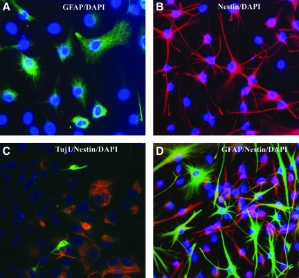
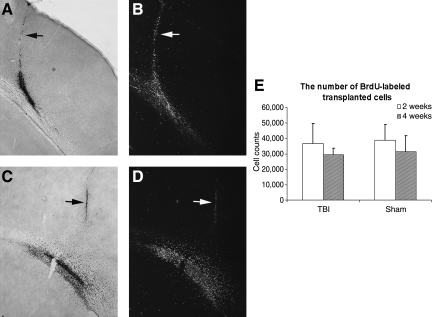
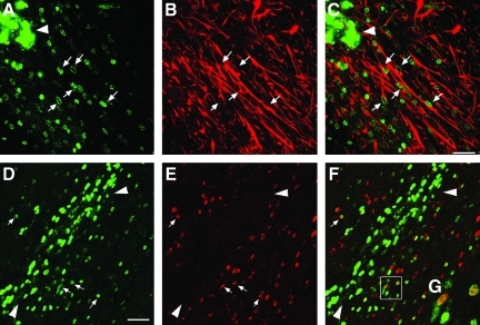
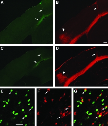
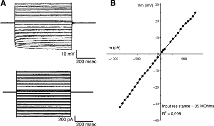
Multipotent neural stem/progenitor cells (NS/NPCs) that are capable of generating neurons and glia offer enormous potential for treating neurological diseases. Adult NS/NPCs that reside in the mature mammalian brain can be isolated and expanded in vitro, and could be a potential source for autologous transplantation to replace cells lost to brain injury or disease. When these cells are transplanted into the normal brain, they can survive and become region-specific cells. However, it has not been reported whether these cells can survive for an extended period and become functional cells in an injured heterotypic environment. In this study, we tested survival, maturation fate, and electrophysiological properties of adult NS/NPCs after transplantation into the injured rat brain. NS/NPCs were isolated from the subventricular zone of adult Fisher 344 rats and cultured as a monolayer. Recipient adult Fisher 344 rats were first subjected to a moderate fluid percussive injury. Two days later, cultured NS/NPCs were injected into the injured brain in an area between the white matter tracts and peri-cortical region directly underneath the injury impact. The animals were sacrificed 2 or 4 weeks after transplantation for immunohistochemical staining or patch-clamp recording. We found that transplanted cells survived well at 2 and 4 weeks. Many cells migrated out of the injection site into surrounding areas expressing astrocyte or oligodendrocyte markers. Whole cell patch-clamp recording at 4 weeks showed that transplanted cells possessed typical mature glial cell properties. These data demonstrate that adult NS/NPCs can survive in an injured heterotypic environment for an extended period and become functional cells.


Figures

FIG. 1.
FIG. 1.
Expanded subventricular zone (SVZ) cells in vitro. After 14 days of in vitro expansion, cultured cells were immunostained with cell nuclear dye 4,6-diamino-2-phenylindole (DAPI, blue) and astrocyte marker glial fibrillary acidic protein (GFAP, A and D, green), progenitor cell marker nestin (B, C, and D, red), and immature neuronal marker Tuj1 (C, green).
FIG. 2.
FIG. 2.
Survival of transplanted cells. Cultured adult rat multipotent neural stem/progenitor cells (NS/NPCs) were labeled with 5-bromo-2-deoxyuridine (BrdU) in vitro 3 days before being used for transplantation and subsequent identification with BrdU immunostaining. (A and B) Coronal section from an animal sacrificed 2 weeks following transplantation. Grafted cells were mostly located at the cortex-white matter interface. (C and D) Coronal section taken from an animal sacrificed at 4 weeks following transplantation. Compared to 2 weeks, at 4 weeks many BrdU-labeled cells had migrated out of the injection site into the surrounding areas. The arrow indicates the injection needle tract. (A and C) Bright-field view. (B and D) Phase-contrast view. (E) Stereological assessment of the number of surviving cells at 2 and 4 weeks following transplantation. The number of cells presented at both time points was similar in sham and injured brains. The total number of BrdU-labeled cells at 4 weeks post-transplantation was lower compared to at 2 weeks in both the sham and injured groups; however, no statistical significance was found (TBI, traumatic brain injury).
FIG. 3.
FIG. 3.
Glial differentiation of transplanted rat multipotent neural stem/progenitor cells (NS/NPCs) in the injured brain at 4 weeks following transplantation. (AC) Astrocytes: confocal micrograph showing double-labeling of 5-bromo-2-deoxyuridine (BrdU; green) and glial fibrillary acidic protein (GFAP, red). Many BrdU-labeled transplanted cells migrated away from the injection center (arrowhead) along the white-matter tract and differentiated into spindle-shaped GFAP-labeled astrocytes (arrows; scale bar=50 μm). (DG) Oligodendrocytes: confocal micrograph showing double-labeling of transplanted cells with BrdU (green) and Olig2 (red). Arrows indicate that many BrdU-positive transplanted cells away from the injection center (arrowhead) were co-labeled with Olig2 (E, red). Merged image shows co-localization of BrdU and Olig2 (F; scale bar=50 μm). (G) Highlighted image of the boxed area in F showing co-labeling of BrdU and Olig2.
FIG. 4.
FIG. 4.
Traumatic brain injury (TBI)- and transplantation-induced focal inflammatory and glial reaction at 4 weeks following cell implantation. (AD) Inflammatory cell reaction as labeled by ED1 staining (B, red) at the cortical injury site (arrowhead), and along the white-matter tract, as well as the cell injection needle tract (arrow). Vimentin-stained glial reaction at the injured cortex, white-matter tract, and the needle tract (D, red). Note 5-bromo-2-deoxyuridine (BrdU)-labeled transplanted cells (A and C, green, arrows) at the site of the peri-cortical-white matter inferface (scale bar=300 μm). (EG) Confocal images of BrdU-labeled transplants (green) co-labeled with ED1 (red). G represents the merged BrdU and ED1 staining (scale bar=40 μm).
FIG. 5.
FIG. 5.
Electrophysiological properties of transplanted multipotent neural stem/progenitor cells (NS/NPCs) at 4 weeks following transplantation. (A) The membrane potential response (top) to current injection (bottom). (B) The amplitude of the membrane potential responses is plotted against the current injected. The input resistance was low and the current-voltage relationship was linear, consistent with mature glial cell but not neuronal electrophysiological properties.
FIG. 6.
FIG. 6.
Cognitive function. The graph compares the Morris water maze (MWM) performance of sham and injured animals receiving injection of vehicle or adult rat multipotent neural stem/progenitor cells (NS/NPCs). Compared to sham animals, injured animals did not show any significant difference in cognitive deficits as assessed by goal latency through days 20–24 following injury. The animals that received cells did not show any difference compared to the vehicle-treated groups (TBI, traumatic brain injury; veh., vehicle).

References

    1. Altman J. Das G.D. Autoradiographic and histological evidence of postnatal hippocampal neurogenesis in rats. J. Comp. Neurol. 1965;124:319–335. - PubMed
    1. Bjorklund A. Kirik D. Rosenblad C. Georgievska B. Lundberg C. Mandel R.J. Towards a neuroprotective gene therapy for Parkinson's disease: use of adenovirus, AAV and lentivirus vectors for gene transfer of GDNF to the nigrostriatal system in the rat Parkinson model. Brain Res. 2000;886:82–98. - PubMed
    1. Boockvar J.A. Schouten J. Royo N. Millard M. Spangler Z. Castelbuono D. Snyder E. O'Rourke D. McIntosh T. Experimental traumatic brain injury modulates the survival, migration, and terminal phenotype of transplanted epidermal growth factor receptor-activated neural stem cells. Neurosurgery. 2005;56:163–171. - PubMed
    1. Dziewczapolski G. Lie D.C. Ray J. Gage F.H. Shults C.W. Survival and differentiation of adult rat-derived neural progenitor cells transplanted to the striatum of hemiparkinsonian rats. Exp. Neurol. 2003;183:653–664. - PubMed
    1. Gage F.H. Coates P.W. Palmer T.D. Kuhn H.G. Fisher L.J. Suhonen J.O. Peterson D.A. Suhr S.T. Ray J. Survival and differentiation of adult neuronal progenitor cells transplanted to the adult brain. Proc. Natl. Acad. Sci. USA. 1995;92:11879–11883. - PMC - PubMed

Publication types

LinkOut - more resources