Review
. 2011:40:143-67.
doi: 10.1146/annurev-biophys-042910-155317.

The cyanobacterial circadian system: from biophysics to bioevolution

Affiliations
Review

The cyanobacterial circadian system: from biophysics to bioevolution

Carl Hirschie Johnson et al. Annu Rev Biophys. 2011.

Abstract

Recent studies have unveiled the molecular machinery responsible for the biological clock in cyanobacteria and found that it exerts pervasive control over cellular processes including global gene expression. Indeed, the entire chromosome undergoes daily cycles of topology/compaction! The circadian system comprises both a posttranslational oscillator (PTO) and a transcriptional/translational feedback loop (TTFL). The PTO can be reconstituted in vitro with three purified proteins (KaiA, KaiB, and KaiC) and ATP. These are the only circadian proteins for which high-resolution structures are available. Phase in this nanoclockwork has been associated with key phosphorylations of KaiC. Structural considerations illuminate the mechanism by which the KaiABC oscillator ratchets unidirectionally. Models of the complete in vivo system have important implications for our understanding of circadian clocks in higher organisms, including mammals. The conjunction of structural, biophysical, and biochemical approaches to this system has brought our understanding of the molecular mechanisms of biological timekeeping to an unprecedented level.


Figures

Figure 1
Figure 1
Circadian rhythms in Synechococcus elongatus. (a) Rhythms of luminescence emanating from cells transformed with bacterial luciferase (luxAluxB) fused to the promoters for the psbAI, purF, kaiBC, ftsZ,and kaiA genes. This plot illustrates circadian rhythms of gene expression. (b) Supercoiling of an endogenous plasmid indicates a circadian rhythm in chromosomal topology. In the subjective night topoisomers of the plasmid are more relaxed (R), whereas in the subjective day they are more supercoiled (SC) (104). (c) Gyrase inhibition results in an immediate change in gene expression due to drug-induced relaxation. Genes that have higher expression during relaxed circadian times immediately increase in gene expression (purF, red), whereas genes that have lower expression during relaxed circadian times immediately decrease in gene expression (kaiC, blue) (from Reference with permission). (d) Micrographs of cyanobacterial cells at different times in constant light. Brightfield images (upper panels) show growth and cell division as a function of approximate circadian time. Observed luminescence (lower panels) reveals circadian rhythms in single cyanobacterial cells. The luminescence reporter was the psbAI promoter driving expression of bacterial luciferase. (e) Quantification of bioluminescence from a single cell as it divides in constant light. Cell division is indicated by differently colored traces for each daughter cell. (Panels d and e courtesy of Dr. Irina Mihalcescu from Reference 55). (f) Cell division in a population of S. elongatus cells is restricted by the circadian system. For the first 36 h the cells are in a light/dark (LD) cycle as indicated by the black and gray bars at the top of the panel. For the remaining time the cells are in constant light (LL). The cell count shows plateaus (red arrows) when the cells stop dividing. Plateaus occur during the night in LD cycles as well as in subjective night of LL. The average doubling time as indicated by the diagonal line was 10.5 h (59).
Figure 2
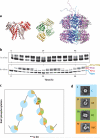
Figure 2
Structures, rhythmic phosphorylation, and associations of KaiA, KaiB, and KaiC. (a) Shown from left to right are the crystal structures of the S. elongatus KaiA dimer (111), the Synechocystis KaiB tetramer (20), and the S. elongatus KaiC hexamer (76, 78). Individual subunits of the multimeric proteins are represented in different colors. In the case of KaiC, the subunits are arranged around a central channel that runs vertically (behind the dark blue-colored subunit in this depiction (13). (b) Time courses of rhythmic KaiC phosphorylation in vivo and in vitro as assessed by SDS-PAGE (the lowest bands are hypophosphorylated KaiC and the upper bands are various forms of phosphorylated KaiC). Top: KaiC phosphorylation in vivo at different times in constant light (samples were collected every 4 h in constant light and immunoblotted). Bottom: KaiC phosphorylation in the in vitro reaction. Purified KaiA, KaiB, and KaiC were combined with ATP in vitro and samples were collected every 3 h and processed for SDS PAGE and staining. Four bands are obvious in these in vitro samples: hypophosphorylated KaiC and KaiC phosphorylated at the S431, T432, or S431/T432 residues (see labels on the right side of the panel). (c) Rhythms of KaiA·KaiB·KaiC complex formation during the in vitro cycling reaction. The color coding of the pie charts indicates the percentage of free KaiC hexamers (blue), KaiA·KaiC complexes (brown), KaiB·KaiC (green) complexes, and KaiA·KaiB·KaiC (orange) complexes (63). (d) Electron microscopy average images of free KaiC hexamers, KaiA·KaiC complexes, KaiB·KaiC complexes, and presumed KaiA·KaiB·KaiC complexes (63) (color coding is the same as in panel c).
Figure 3
Figure 3
Models of the KaiABC oscillator. (a) Diagram representing the mathematical model for the KaiC phosphorylation cycle. The double circle dumbbell shapes in the center represent KaiC monomers. The KaiC hexamer can associate with and dissociate from KaiA and KaiB. KaiC hexamers are shown in light blue and dark blue, representing two conformational states (approximately equivalent to kinase versus phosphatase forms of KaiC). Red dots are phosphates at KaiC phosphorylation sites (residues S431 and T432). Monomer exchange between KaiC hexamers is depicted with the double-headed arrows in the center. The rates of monomer exchange vary among KaiC states, with a solid line indicating a high rate and a dashed line indicating a low rate (from Reference 63). (b) Diagram showing the formation of KaiA·KaiB·KaiC complexes. Starting from the leftmost molecular representation and proceeding clockwise: during the phosphorylation phase of the cycling reaction, KaiA (red dimers) repeatedly and rapidly interacts with KaiC's C-terminal tentacles. (KaiC molecules are the green double-donut hexamers.) When KaiC becomes hyperphosphorylated (phosphates on T432 and S431 are depicted as red dots), it first binds KaiB (green diamonds) stably. Then, the KaiB·KaiC complex binds KaiA, sequestering it from further interaction with KaiC's tentacles. At that point, KaiC initiates dephosphorylation. When KaiC is hypophosphorylated, it releases KaiB and KaiA, thereby launching a new cycle (from Reference 81).
Figure 4
Figure 4
A ratcheting mechanism for unidirectional motion of the KaiABC oscillator. Starting with the unphosphorylated (unphos) form of KaiC (S/T), KaiC is first phosphorylated on T432 (S/pT), leading to the formation of a salt bridge (solid green line) to R385 on the adjacent KaiC subunit (blue chain; T432-P shows proximity to E318, as shown by the dashed green line). KaiC then autophosphorylates on S431, leading to the doubly phosphorylated form (pS/pT) that adds hydrogen bonds (solid green lines) to residues T426 and H429 on the same KaiC subunit (pink chain). The formation of these hydrogen bonds makes the reverse reactions unfavorable so that the KaiABC oscillator is unidirectional during the phosphorylation phase (33). The hyperphosphorylated KaiC (pS/pT) then interacts with KaiB and initiates monomer exchange and dephosphorylation, forming first pS/T and ultimately unphosphorylated KaiC (S/T) again. The S/T and pS/T forms of KaiC are inferred and labeled as hypothetical models on the figure because no crystal structures of these forms have been reported, whereas the S/pT and pS/pT forms have been successfully crystallized and reported (76, 75, 108).
Figure 5
Figure 5
FRET analysis of KaiC monomer exchange. (a) A sample of KaiC labeled with IAEDANS (EX 336/EM 470 nm) was mixed with a sample of KaiC labeled with MTSF (EX 490/EM 515 nm). The emission spectrum of the mixture under excitation at 336 nm was recorded at the following times at 30°C: 0 h, 0.16 h, 0.5 h, 1 h, 2 h, 4 h, 6 h, and 8 h. The decrease in fluorescence intensity at 470 nm of IAEDANS-labeled KaiC is indicative of energy transfer due to monomer exchange between the two labeled KaiC populations. (b) Effect of KaiA and KaiB on monomer exchange. Measurement of monomer exchange between IAEDANS-labeled and MTSF-labeled KaiC when KaiA (0.05 μg μl–1) or KaiB (0.05 μg μl–1) was added to the mixture of KaiC (0.2 μg μl–1 total concentration). The decrease in fluorescence intensity at 470 nm was plotted as a function of time. (c) Model prediction of the in vitro KaiABC oscillation in the presence (blue line) or absence (red line) of phase-dependent monomer exchange (from Reference 63). Abbreviations: FRET, fluorescence resonance energy transfer; IAEDANS, 5-((((2-iodoacetyl)amino)ethyl)amino)naphthalene-1-sulfonic acid; MTSF, 2-((5-fluoresceinyl)aminocarbonyl)ethyl methanethiosulfonate-4-fluorescein.
Figure 6
Figure 6
The core posttranslational oscillator (PTO) is embedded in a larger transcription/translation feedback loop (TTFL). The PTO is linked to the damped TTFL by transcription and translation of the kaiABC cluster. Global gene expression is mediated by rhythmic modulation of the activity of all promoters, including those driving the expression of the central clock gene cluster, kaiABC (ABC in figure). Rhythmic DNA torsion and/or transcriptional factor activity (e.g., SasA/RpaA) modulates global promoter activities. Cyclic changes in the phosphorylation status of KaiC regulate DNA topology/transcriptional factors. The PTO is determined by KaiC phosphorylation as regulated by interactions with KaiA and KaiB (compare with Figure 3a). Robustness is maintained by synchronization of KaiC hexameric status via monomer exchange (depicted by dumbbell-shaped KaiC monomers exchanging with KaiC hexamers in the middle of the PTO cycle). The shade of KaiC hexamers (dark versus light blue) denotes conformational changes that roughly equate to kinase versus phosphatase forms. New synthesis of KaiC feeds into the KaiABC oscillator as nonphosphorylated hexamers or as monomers that exchange into pre-existing hexamers. If the new synthesis of KaiC occurs at a phase when hexamers are predominantly hypophosphorylated, the oscillation of KaiC phosphorylation is reinforced (enhanced amplitude). If new synthesis of unphosphorylated KaiC happens at a phase when hexamers are predominantly hyperphosphorylated, this leads to an overall decrease in the KaiC phosphorylation status, thereby altering the phase of the KaiABC oscillator (phase shift), reducing its amplitude, or both. Phase shifts accomplished by this mechanism could be partially or totally responsible for entrainment in vivo (from Reference 82).

References

    1. Akiyama S, Nohara A, Ito K, Maéda Y. Assembly and disassembly dynamics of the cyanobacterial periodosome. Mol. Cell. 2008;29:703–16. - PubMed
    1. Amdaoud M, Vallade M, Weiss-Schaber C, Mihalcescu I. Cyanobacterial clock, a stable phase oscillator with negligible intercellular coupling. Proc. Natl. Acad. Sci. USA. 2007;104:7051–56. - PMC - PubMed
    1. Andersson CR, Tsinoremas NF, Shelton J, Lebedeva NV, Yarrow J, et al. Application of bioluminescence to the study of circadian rhythms in cyanobacteria. Methods Enzymol. 2000;305:527–42. - PubMed
    1. Angermayr SA, Hellingwerf KJ, Lindblad P, de Mattos MJ. Energy biotechnology with cyanobacteria. Curr. Opin. Biotechnol. 2009;20:257–63. - PubMed
    1. Atsumi S, Higashide W, Liao JC. Direct photosynthetic recycling of carbon dioxide to isobutyraldehyde. Nat. Biotechnol. 2009;27:1177–80. - PubMed

Publication types

MeSH terms

Substances

LinkOut - more resources