. 2011 Aug;10(4):584-94.
doi: 10.1111/j.1474-9726.2011.00686.x. Epub 2011 Apr 7.

Mitochondrial peptidase IMMP2L mutation causes early onset of age-associated disorders and impairs adult stem cell self-renewal

Affiliations

Mitochondrial peptidase IMMP2L mutation causes early onset of age-associated disorders and impairs adult stem cell self-renewal

Sunil K George et al. Aging Cell. 2011 Aug.

Abstract

Mitochondrial reactive oxygen species (ROS) are proposed to play a central role in aging and age-associated disorders, although direct in vivo evidence is lacking. We recently generated a mouse mutant with mutated inner mitochondrial membrane peptidase 2-like (Immp2l) gene, which impairs the signal peptide sequence processing of mitochondrial proteins cytochrome c1 and glycerol phosphate dehydrogenase 2. The mitochondria from mutant mice generate elevated levels of superoxide ion and cause impaired fertility in both sexes. Here, we design experiments to examine the effects of excessive mitochondrial ROS generation on health span. We show that Immp2l mutation increases oxidative stress in multiple organs such as the brain and the kidney, although expression of superoxide dismutases in these tissues of the mutants is also increased. The mutants show multiple aging-associated phenotypes, including wasting, sarcopenia, loss of subcutaneous fat, kyphosis, and ataxia, with female mutants showing earlier onset and more severe age-associated disorders than male mutants. The loss of body weight and fat was unrelated to food intake. Adipose-derived stromal cells (ADSC) from mutant mice showed impaired proliferation capability, formed significantly less and smaller colonies in colony formation assays, although they retained adipogenic differentiation capability in vitro. This functional impairment was accompanied by increased levels of oxidative stress. Our data showed that mitochondrial ROS is the driving force of accelerated aging and suggested that ROS damage to adult stem cells could be one of the mechanisms for age-associated disorders.


Figures

Fig. 1
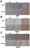
Fig. 1. Elevated expression of 4-HNE-protein adducts and SOD in mutant tissues
A. HNE-protein adducts expression was significantly elevated in the brain and kidney of mutant mice. B. SOD1 expression was increased in the brain and kidney of mutant mice. C. Increased SOD2 expression in the brain and kidney of mutant mice. Brain and kidney tissues were from females of 16 months. Positive signals were visualized by 3-amino-9-ethylcarbazole, which stained reddish-brown. Nuclei were counterstained with hematoxylin. +/+, +/− and −/− indicate normal control, heterozygous and homozygous mutant mice. Shown were typical staining results for tissues from at least 3 animals of each genotype.
Fig. 2
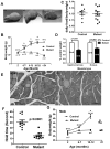
Fig. 2. Aging-associated phenotypes in mutant mice
A. Kyphosis of 24-month-old female mutant mice. Note the difference of coat color due to different expression of transgenic tyrosinase in heterozygous (+/−) and mutant (−/−) mice. B. Reduced bodyweight of aged mutant females. Number of animals of each time point is listed. Control mice include +/+ and +/− genotypes. * indicates significant difference by ANOVA. Animals beyond 24 months were excluded from ANOVA analysis due to limited sample number. C. No difference in food consumption between 6–7 months female control and mutant mice. D. Reduced skeletal muscle mass of 20–22 month mutant females. Numbers of samples are listed in the columns. Since each time one mutant and one age-matched control were sacrificed at the same time, muscle mass from the mutant was expressed as fraction of that of the control. Significant difference was found between control and mutants by ANOVA. E. H&E staining of soleus muscle from 21-month females. F. Wire hang assay showed reduced grip strength of mutant mice at 21 months. G. Bodyweight of mutant males. Significant difference can only be observed at 10–12 months (ANOVA). Available bodyweight data beyond 10–12 months are also presented in the figure but these data are excluded from ANOVA analysis due to the limited number of samples. * indicates significant difference by ANOVA. Means ±s.e.m. are represented in B, C, D, E and G.
Fig. 3
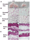
Fig. 3. Loss of adipose tissue in aged mutant females
A. Smaller gonadal adipose tissue of 10-month mutant females. B. H&E staining of gonadal fat tissue of 10-month females. No difference in size of adipocytes is noted between control and mutants. C. H&E staining of perirenal fat tissue of 24-month females. No difference in size of adipocytes is noted between control and mutants. D. E. H&E staining of skin cross section showing the loss of subcutaneous fat in 24-months but not 4-months mutant females. Scale bars: 50 μ.
Fig. 4
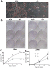
Fig. 4. Adult stem cell defects in mutant animals
A. Adipogenesis was impaired in adipose-derived stromal cells (ADSCs) from mutant mice. Mature adipocytes were stained with Oil Red O. B. ADSC cells from mutant mice formed fewer and smaller colonies in colony formation assay. Giemsa staining was performed to help visualize the cells. Differences were observed in ADSCs from gonadal adipose tissue of 10-month-old mice, but were more pronounced in ADSCs from gonadal adipose tissue of 24-month-old mice. C. Proliferation assays for ADSC cells from control and mutant mice. D. Proliferation assays for bone marrow-derived stromal cells from control and mutant mice. For C and D, triplicate assays were performed for each experiment. The experiments were repeated at least three times and similar results were observed. Data from one experiment are shown; mean±s.e.m..*, p<0.05.
Fig. 5
Fig. 5. Elevated oxidative stress in adipose-derived stromal cells from mutant mice
A. Elevated expression of HNE-protein adducts, superoxide dismutase 1 and 2 in mutant adipose-derived stromal cells. B. Lower GSH concentrations in mutant adipose-derived stromal cells. *, p<0.05.

Similar articles

Cited by

References

    1. Bayne AC, Mockett RJ, Orr WC, Sohal RS. Enhanced catabolism of mitochondrial superoxide/hydrogen peroxide and aging in transgenic Drosophila. Biochem J. 2005;391:277–284. - PMC - PubMed
    1. Behrens M, Michaelis G, Pratje E. Mitochondrial inner membrane protease 1 of Saccharomyces cerevisiae shows sequence similarity to the Escherichia coli leader peptidase. Mol Gen Genet. 1991;228:167–176. - PubMed
    1. Brand MD. The sites and topology of mitochondrial superoxide production. Exp Gerontol. 2010;45:466–472. - PMC - PubMed
    1. Caplan AI. Adult mesenchymal stem cells for tissue engineering versus regenerative medicine. Journal of Cellular Physiology. 2007;213:341–347. - PubMed
    1. Chehab O, Ouertani M, Souiden Y, Chaieb K, Mahdouani K. Plasma antioxidants and human aging: a study on healthy elderly Tunisian population. Mol Biotechnol. 2008;40:27–37. - PubMed

MeSH terms

Substances