. 2011 May;90(5):376-89.
doi: 10.1016/j.ejcb.2010.11.016. Epub 2011 Feb 18.

AFAP1L1 is a novel adaptor protein of the AFAP family that interacts with cortactin and localizes to invadosomes

Affiliations

AFAP1L1 is a novel adaptor protein of the AFAP family that interacts with cortactin and localizes to invadosomes

Brandi N Snyder et al. Eur J Cell Biol. 2011 May.

Abstract

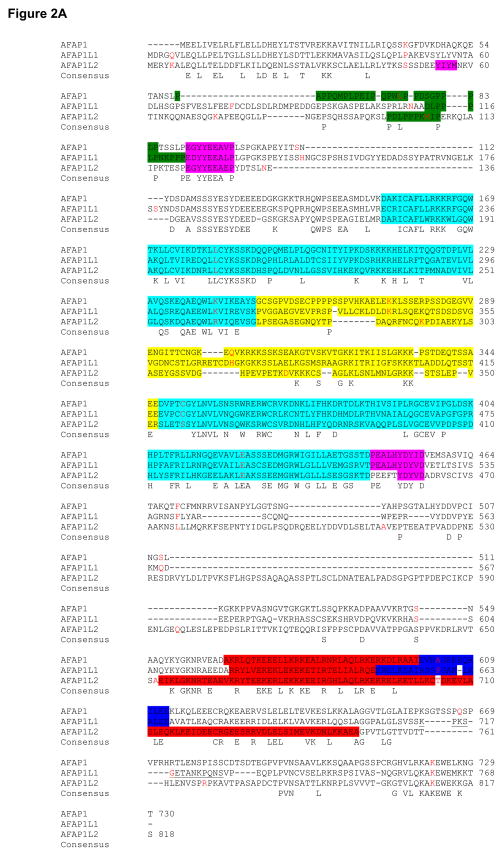
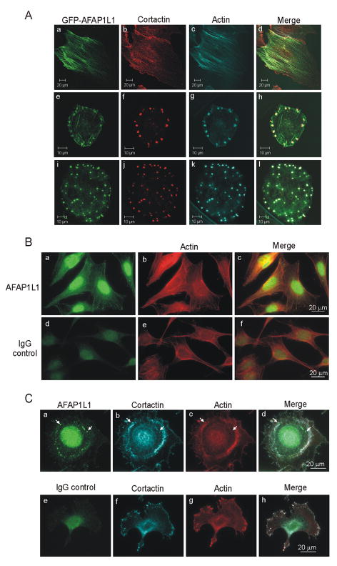
The actin-filament associated protein (AFAP) family of adaptor proteins consists of three members: AFAP1, AFAP1L1, and AFAP1L2/XB130 with AFAP1 being the best described as a cSrc binding partner and actin cross-linking protein. A homology search of AFAP1 recently identified AFAP1L1 which has a similar sequence, domain structure and cellular localization; however, based upon sequence variations, AFAP1L1 is hypothesized to have unique functions that are distinct from AFAP1. While AFAP1 has the ability to bind to the SH3 domain of the nonreceptor tyrosine kinase cSrc via an N-terminal SH3 binding motif, it was unable to bind cortactin. However, the SH3 binding motif of AFAP1L1 was more efficient at interacting with the SH3 domain of cortactin and not cSrc. AFAP1L1 was shown by fluorescence microscopy to decorate actin filaments and move to punctate actin structures and colocalize with cortactin, consistent with localization to invadosomes. Upon overexpression in A7r5 cells, AFAP1L1 had the ability to induce podosome formation and move to podosomes without stimulation. Immunohistochemical analysis of AFAP1L1 in human tissues shows differential expression when contrasted with AFAP1 with localization of AFAP1L1 to unique sites in muscle and the dentate nucleus of the brain where AFAP1 was not detectable. We hypothesize AFAP1L1 may play a similar role to AFAP1 in affecting changes in actin filaments and bridging interactions with binding partners, but we hypothesize that AFAP1L1 may forge unique protein interactions in which AFAP1 is less efficient, and these interactions may allow AFAP1L1 to affect invadosome formation.


Figures

Figure 1
Figure 1. AFAP1L1 sequence
AFAP1L1 coding sequence was divided into codons with corresponding amino acid sequence. Start and stop codons are indicated in bold type.
Figure 2
Figure 2. AFAP family members share both sequence and domain similarity
A. AFAP1, AFAP1L1 and AFAP1L2 amino acid sequences were compared using ClustalW2 alignment (Larkin et al., 2007). Consensus sequence between all three family members is labeled as consensus. Intron/exon boundaries are marked by red letters. Predicted SH3 binding motifs are highlighted in green, predicted SH2 binding motifs in pink, predicted PH domains in light blue, predicted Substrate Domain (SD) in yellow, predicted leucine zipper (AFAP1, AFAP1L1) and coiled coil (AFAP1L2) in red and predicted actin binding domain in dark blue. The AFAP1L1 peptide sequence used to create 1L1-CT antibody is underlined. B. Modular domain organization of AFAP family members was compared. SH3bm = SH3 binding motif, SH2bm = SH2 binding motif, PH1 = pleckstrin homology domain 1, PH2 = pleckstrin homology domain 2, SD = serine/threonine rich substrate domain, Lzip = leucine zipper, ABD = actin binding domain. Sequences that do not correlate with an identified type of modular domain or motif are labeled “A, B, C, D or E”.
Figure 3
Figure 3. A novel antibody specifically recognizes AFAP1L1
A. Cell lines were lysed in 2X SDS buffer, resolved by 8% SDS-PAGE and transferred to PVDF. The Sigma antibody Ab2 specifically recognized a protein band of 115 kDa. Bands identified as AFAP1L1 and recognized by the Ab2 antibody (top panel) are indicated by an arrow. Gamma tubulin was used as a loading control. Not shown, but in an adjacent lane, was a lysate prepared from 293T cells transfected with untagged AFAP1L1 which was used in identifying the band corresponding to AFAP1L1. B. Endogenous AFAP1L1 and AFAP1 were specifically immunoprecipitated from Cos-1 cells using 5μg 1L1-CT and F1 polyclonal antibodies respectively and the resolved proteins detected with 1L1-CT (left panel) or AFAP1 monoclonal antibodies (BD Transduction, right panel). Rabbit IgG antibody was used as a control. Note the differences in the molecular weight markers for each western. C. GFP-AFAP1L1 and GFP-AFAP1 were overexpressed in Cos-1 cells and lysed in 2X SDS buffer. Lysates were resolved by 8% SDS-PAGE, transferred to PVDF membrane and immunoblotted with either AFAP1L1 (1L1-CT) or AFAP1 (F1) antibody. GFP (top right western) and β-actin (bottom westerns) were used as loading controls. D. The peptide sequence used to create antibody 1L1-CT was compared to analogous sequences in human, chimpanzee, mouse and rat to show similarity between antibody binding sites.
Figure 4
Figure 4. Immunohistochemical analysis of AFAP1L1 shows differential expression from AFAP1 in human tissue
Paraffin-embedded human breast (A), colon (B) and brain (C) tissues were analyzed for AFAP1 and AFAP1L1 localization using F1 and 1L1-CT antibodies respectively. Breast regions include breast ducts (4A panels a–d), breast lobules (4A panels a,b) and microvasculature (4A panels e,f). Colon regions include the mucosa (4B panels a,b,g,h), lamina propria (4B panels a–d, g,h) and muscularis (4B panels a–f). Brain regions include the cerebellar cortex (4C panels a–j), dentate nucleus (4C panels k–n) and glial cells (4C panels o,p). AFAP1L1 is designated by long thin arrows while AFAP1 is designated by arrowheads.
Figure 5
Figure 5. Subcellular localization of GFP-AFAP1L1 shows association with actin and invadosomes
A. A7r5 cells transiently expressing GFP-AFAP1L1 were plated onto fibronectin-coated coverslips and immunolabeled for cortactin (Millipore). Actin was visualized with TRITC-phalloidin (Sigma). Representative images of cells with well-formed stress fibers or podosome formation are shown. B. MDA-MB-435 cells were plated on fibronectin coated coverslips and immunolabeled for endogenous AFAP1L1 (Sigma Ab2, panel a–c). Rabbit IgG was used as a control (panel d–f) and actin was visualized with AlexaFluor labeled phalloidin. Epifluorescence images of representative cells are shown. C. MDA-MB-435 cells were transfected with Src 527F construct, plated onto fibronectin coated coverslips and immunolabeled for AFAP1L1 (Sigma Ab1, panel a–d). Rabbit IgG was used as control antisera for AFAP1L1 antibody (panel e–h). Cortactin was immunolabeled with monoclonal anti-cortactin antibodies (4F11, Millipore) and actin was visualized by AlexaFluor labeled phalloidin (panel c, g). Examples of AFAP1L1 co-localizing to invadopodia, actin and cortactin rich punctate structures are marked with white arrows.
Figure 6
Figure 6. Podosome formation in A7r5 transfected with GFP-AFAP1 or GFP-AFAP1L1 plasmids
A7r5 cells were transfected with the indicated amounts of plasmids encoding either GFP-AFAP1 or GFP-AFAP1L1 (in combination with pcDNA3.1) to bring the total amount of plasmid DNA to 1 μg for each transfection. Twenty four hours post-transfection, the cells were transferred to fibronectin-coated coverslips. Forty-eight hours after transfection, cells were processed for immunofluorescence analysis or used to prepare whole cell SDS lysates. (A) Equal amounts of SDS lysates were resolved by 8% SDS-PAGE and then transferred to PVDF membranes and subsequently probed with an antiserum that recognizes GFP. (B) Cells expressing GFP-AFAP1 or GFP-AFAP1L1 were assessed for podosome formation and the percentage of cells exhibiting podosomes for each transfection was calculated. 150 to 300 cells were counted for each transfection. Panel A and B represents one experiment out of two independently performed experiments.
Figure 7
Figure 7. AFAP1L1 interacts with cortactin SH3 domain
A. 1 mg of lysate from 293T cells transiently expressing GFP-AFAP1 was incubated with 50 μg of GST, GST-Src-SH3 domain or GST-cortactin-SH3 domain bound fusion protein to glutathione Sepharose 4B beads and probed for GFP through western blot analysis (upper panel). The lower panel represents a GelCode Blue Stain (ThermoScientific) of GST or GST fusion protein. A graph representing the ratio of AFAP1 pulled down by each GST fusion protein compared to GST control using scanning densitometry is shown. B. 1mg of lysate from 293T cells transiently expressing GFP-AFAP1L1 was incubated with 50μg of GST, GST-Src-SH3 domain or GST-cortactin-SH3 domain bound to glutathione Sepharose 4B beads and probed for GFP through western blot analysis (upper panel). The lower panel represents a GelCode Blue Stain (ThermoScientific) of GST or GST fusion protein. A graph representing the ratio of AFAP1L1 pulled down by each GST fusion protein compared to GST control using scanning densitometry is shown. C. 293T transiently expressing cortactin with either GFP-AFAP1 or GFP-AFAP1L1 were immunoprecipitated with anti-GFP antibody, probed for cortactin through western blot analysis (upper panel) and then re-probed for GFP tagged proteins (lower panel). D. 293T transiently expressing cortactin with either GFP-AFAP1 or GFP-AFAP1L1 were immunoprecipitated with anti-cortactin antibody, probed for GFP tagged proteins through western blot analysis (upper panel) and then re-probed for cortactin (lower panel).

References

    1. Baisden JM, Gatesman AS, Cherezova L, Jiang BH, Flynn DC. The intrinsic ability of AFAP-110 to alter actin filament integrity is linked with its ability to also activate cellular tyrosine kinases. Oncogene. 2001a;20:6607–6616. - PubMed
    1. Baisden JM, Qian Y, Zot HM, Flynn DC. The actin filament-associated protein AFAP-110 is an adaptor protein that modulates changes in actin filament integrity. Oncogene. 2001b;20:6435–6447. - PubMed
    1. Braak E, Arai K, Braak H. Cerebellar involvement in Pick’s disease: affliction of mossy fibers, monodendritic brush cells, and dentate projection neurons. Exp Neurol. 1999;159:153–163. - PubMed
    1. DiNitto JP, Lambright DG. Membrane and juxtamembrane targeting by PH and PTB domains. Biochim Biophys Acta. 2006;1761:850–867. - PubMed
    1. Dorfleutner A, Cho Y, Vincent D, Cunnick J, Lin H, Weed SA, Stehlik C, Flynn DC. Phosphorylation of AFAP-110 affects podosome lifespan in A7r5 cells. J Cell Sci. 2008;121:2394–2405. - PubMed

Publication types

LinkOut - more resources