. 2011 Mar 8;21(5):345-52.
doi: 10.1016/j.cub.2011.01.039. Epub 2011 Feb 17.

Plant stem cell signaling involves ligand-dependent trafficking of the CLAVATA1 receptor kinase

Affiliations

Plant stem cell signaling involves ligand-dependent trafficking of the CLAVATA1 receptor kinase

Zachary L Nimchuk et al. Curr Biol. .

Abstract

Background: Cell numbers in above-ground meristems of plants are thought to be maintained by a feedback loop driven by perception of the glycopeptide ligand CLAVATA3 (CLV3) by the CLAVATA1 (CLV1) receptor kinase and the CLV2/CORYNE (CRN) receptor-like complex. CLV3 produced in the stem cells at the meristem apex limits the expression level of the stem cell-promoting homeodomain protein WUSCHEL (WUS) in the cells beneath, where CLV1 and WUS RNA are localized. WUS downregulation nonautonomously reduces stem cell proliferation. Overexpression of CLV3 eliminates the stem cells, causing meristem termination, and loss of CLV3 function allows meristem overproliferation. There are many questions regarding the CLV3/CLV1 interaction, including where in the meristem it occurs, how it is regulated, and how it is that a large range of CLV3 concentrations gives no meristem size phenotype.

Results: Here we use genetics and live imaging to examine the cell biology of CLV1 in Arabidopsis meristematic tissue. We demonstrate that plasma membrane-localized CLV1 is reduced in concentration by CLV3, which causes trafficking of CLV1 to lytic vacuoles. We find that changes in CLV2 activity have no detectable effects on CLV1 levels. We also find that CLV3 appears to diffuse broadly in meristems, contrary to a recent sequestration model.

Conclusions: This study provides a new model for CLV1 function in plant stem cell maintenance and suggests that downregulation of plasma membrane-localized CLV1 by its CLV3 ligand can account for the buffering of CLV3 signaling in the maintenance of stem cell pools in plants.


Figures

Figure 1
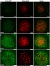
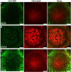
Figure 1. CLV1 accumulates at the plasma membrane in clv3-2 plants
Images represent single clv1-11 CLV1∷CLV1-2xGFP transgenic line introgressed into clv1-11 clv3-2 plants. Images are of L3/L4 cell layer of young IMs (see text). A and B rows are an average of four scans. C and D rows are a sum of four scans to highlight GFP signal. See Supplemental figures for imaging controls. Scale bar, 10 μm.
Figure 2
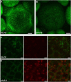
Figure 2. CLV1 traffics to the vacuole in a CLV3-dependent manner
Images from an introgression of a single pUBQ∷CLV1-2xGFP transgenic line in the clv1-101 background into clv1-11 clv3-2 plants (see text). Images are of A) CLV3 ER and B) clv3-2 ER plants. Images represent reconstructed stack of young IMs. Scale bar, 20 μm. C and D, detail images from a separate introgression of a different UBQ∷CLV1-2xGFP transgenic line in the clv1-11 er background into the genotype clv1-11 clv3-2 er. Images are of the L1 epidermal cell layer and represent and average of four scans. Scale bars, 2 μm.
Figure 3
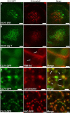
Figure 3. CLV1 traffics to the lytic vacuole in a VTI11 dependent manner
Images of L3 cells of young IMs from an introgression of pCLV1∷CLV1-2xGFP clv1-11 into the clv1-11 clv3-2 zig-1 triple mutant background (see text). Row A: Scan of L3 cells from IM of pCLV1∷CLV1-2xGFP CLV3 ZIG. Row B: Scan of L3 cells from pCLV1∷CLV1-2xGFP CLV3 zig-1. Images are a sum of 4 scans. Scale bars, 10 μm. Row C: Confocal image of IM L1 cells from pUBQ∷CLV1-2xGFP CLV3 plant stained for 3 hrs with FM4-64. Arrows indicate FM4-64 localization to the tonoplast surrounding the CLV1-2xGFP signal. Images are an average of 4 scans. Scale Bars, 1 mm. Row D: Confocal image of IM L1 cells from pUBQ∷CLV1-2xGFP CLV3 plant stained for 5 minutes with LysoTracker Red. Arrows indicate areas of colocalization between CLV1-2xGFP and LysoTracker Red in vacuoles. Images are an average of 4 scans. Scale Bars, 2 2 μm. Row E: Confocal image of IM L1 cells from pUBQ∷CLV1-2xGFP VAC-RFP CLV3 plants. Scale Bars, 2 2 μm. Images are an average of 2 scans.
Figure 4
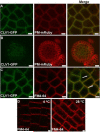
Figure 4. CLV1 localizes to the plasma membrane in clv3 plants
Row A: CLV1-2xGFP colocalizes with PM-mRUBY in clv3-2 plants. Confocal image of IM L1 cells from pUBQ∷CLV1-2xGFP pUBQ:PM-2xmRUBY clv3-2. Images are an average of 4 scans. Scale bars, 10 μm. Row B: CLV1-2xGFP expressed from the pCLV1∷CLV1-2xGFP transgene in a pUBQ:PM-2xmRUBY clv3-2 FM colocalizes with PM-2xmRUBY. Images are an average of 4 scans. Scale bars, 10 μm.. Row C: CLV1 colocalizes with FM4-64 at the PM in cold treated IMs. Arrows indicate CLV1-GFP vesicles lacking FM4-64 signal. Row D: Cold treatment abolishes FM4-64 endocytosis in root cells. Seedling roots were incubated for 1 hr with FM4-64 at 4°C or 20 minutes at 28°C. Note the lack of FM4-64 positive vesicles at 4°C.
Figure 5
Figure 5. High levels of CLV1 in L1 and L2 fail to prevent CLV3-dependent trafficking of CLV1 in L3 cells
A and B, images taken from L3 cells of young IMs from the clv1-11 UBQ∷CLV1-2xGFP introgression into the clv3-2 clv1-11 background. A and B represent an average of four scans. C, a sum of four scans from CLV3 progeny to highlight weak signals in the L3 cells.
Figure 6
Figure 6. CLV3 is sufficient to drive CLV1 downregulation at the plasma membrane and trafficking to the lytic vacuole
Inducible expression of CLV3 restores vacuolar trafficking of CLV1-2xGFP. Images are derived from F1 progeny of a cross between clv3-2 DEX∷CLV3 and clv3-2 UBQ∷CLV1-2xGFP plants. A, Image taken from young IMs from an F1 plant before application of dexamethasone (DEX). Note the robust accumulation of CLV1-2xGFP on the PM. B, different IM from the same F1 plant following 3 days of spraying with 20 μm DEX. Note the strong vacuolar accumulation of GFP signal. C, a different IM from the same DEX treated plant displaying greatly reduced levels of CLV1-2xGFP at the PM. D, an IM taken from a control treated F1 plant at the same time as B and C. Images represent a sum of 4 scans. Scale bars, 5 μm.
Figure 7
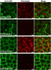
Figure 7. CLV2 is not required for CLV1 trafficking or stability
CLV1-2xGFP traffics to the lytic vacuole independent of CLV2. Image of L1 tissue from pUBQ∷CLV1-2xGFP plants introgressed into the CLV2 (upper) or clv2-1 (lower) background. Image represents average of four scans. Scale bars, 2 μm. B). CLV1-2xGFP stability in clv3-2 plants is not compromised in clv2-1 plants. Image of L1 cells in young IMs from an introgression of pUBQ∷CLV1-2xGFP into the clv3-2 CLV2 (upper) or clv3-2 clv2-1 (lower) backgrounds. Image represents average of four scans. Scale bars, 2 μm.

References

    1. Wang G, Fiers M. CLE peptide signaling during plant development. Protoplasma. 2009;240:33–43. - PMC - PubMed
    1. Brand U, Fletcher JC, Hobe M, Meyerowitz EM, Simon R. Dependence of stem cell fate in Arabidopsis on a feedback loop regulated by CLV3 activity. Science. 2000;289:617–619. - PubMed
    1. Clark SE, Running MP, Meyerowitz EM. CLAVATA3 is a specific regulator of shoot and floral meristem development affecting the same processes as CLAVATA1. Development. 1995;121:2057–2067.
    1. Muller R, Borghi L, Kwiatkowska D, Laufs P, Simon R. Dynamic and compensatory responses of Arabidopsis shoot and floral meristems to CLV3 signaling. Plant Cell. 2006;18:1188–1198. - PMC - PubMed
    1. Lenhard M, Laux T. Stem cell homeostasis in the Arabidopsis shoot meristem is regulated by intercellular movement of CLAVATA3 and its sequestration by CLAVATA1. Development. 2003;130:3163–3173. - PubMed

Publication types

MeSH terms

Substances