

Oxytocin antagonist disrupts male mouse medial amygdala response to chemical-communication signals
- PMID: 21333718
- PMCID: PMC3093756
- DOI: 10.1016/j.neuroscience.2011.02.030
Oxytocin antagonist disrupts male mouse medial amygdala response to chemical-communication signals
Abstract
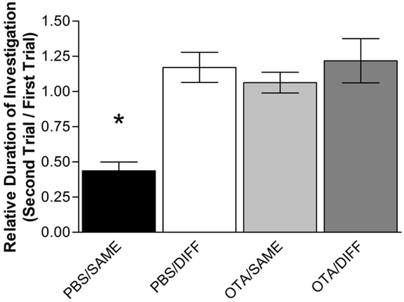
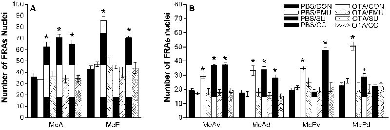
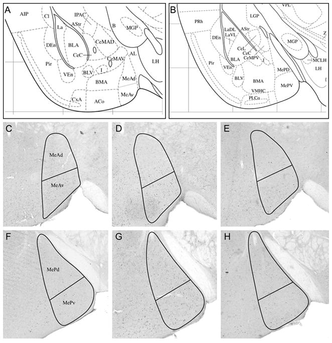
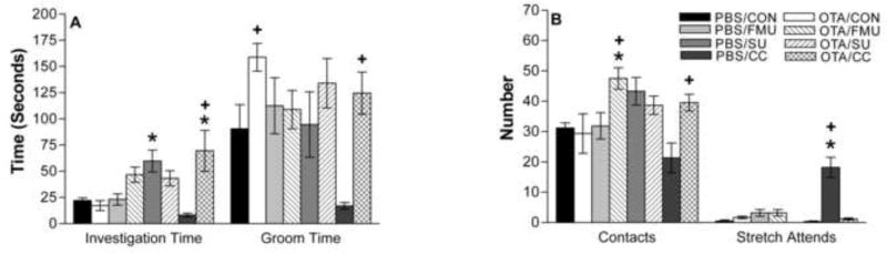
The male mouse medial amygdala is an important site for integration of main and accessory olfactory information. Exposure to biologically relevant chemical signals from the same species (conspecific) results in a general pattern of immediate early gene (IEG) expression in medial amygdala different from that elicited by chemical signals from other species (heterospecific), of no demonstrable biological relevance. The neuropeptide oxytocin (OT) in the medial amygdala has been shown to be necessary for social recognition. In the present set of experiments, male mice with i.c.v. cannulae were injected with either PBS (vehicle control) or oxytocin antagonist (OTA) (1 ng in 1 μl PBS) and exposed to conspecific (female mouse urine) and heterospecific (steer urine and worn cat collar) chemical stimuli. Similarly to our previous report with intact male mice [Samuelsen and Meredith (2009a) Brain Res 1263:33-42], PBS-injected mice exhibited different immediate early gene (IEG) expression patterns in the medial amygdala according to the biological relevance of the chemical stimuli. However, OTA injection eliminates the increase in IEG expression in the medial amygdala to any of the tested conspecific or heterospecific stimuli. Importantly, OTA injection disrupts avoidance of an unfamiliar predator odor, worn cat collar. Here we suggest that the disruption of social recognition behavior in male mice with altered OT receptor activity results from an inability of the medial amygdala to process relevant conspecific (and heterospecific) chemosensory signals.
Copyright © 2011 IBRO. Published by Elsevier Ltd. All rights reserved.
Figures
Similar articles
-
The vomeronasal organ is required for the male mouse medial amygdala response to chemical-communication signals, as assessed by immediate early gene expression.Neuroscience. 2009 Dec 29;164(4):1468-76. doi: 10.1016/j.neuroscience.2009.09.030. Epub 2009 Sep 22. Neuroscience. 2009. PMID: 19778594 Free PMC article.
-
Categorization of biologically relevant chemical signals in the medial amygdala.Brain Res. 2009 Mar 31;1263:33-42. doi: 10.1016/j.brainres.2009.01.048. Epub 2009 Feb 5. Brain Res. 2009. PMID: 19368822 Free PMC article.
-
Characteristic Response to Chemosensory Signals in GABAergic Cells of Medial Amygdala Is Not Driven by Main Olfactory Input.Chem Senses. 2017 Jan;42(1):13-24. doi: 10.1093/chemse/bjw096. Epub 2016 Sep 20. Chem Senses. 2017. PMID: 27651427 Free PMC article.
-
Influence of Cat Odor on Reproductive Behavior and Physiology in the House Mouse: (Mus Musculus).In: Mucignat-Caretta C, editor. Neurobiology of Chemical Communication. Boca Raton (FL): CRC Press/Taylor & Francis; 2014. Chapter 14. In: Mucignat-Caretta C, editor. Neurobiology of Chemical Communication. Boca Raton (FL): CRC Press/Taylor & Francis; 2014. Chapter 14. PMID: 24830030 Free Books & Documents. Review.
-
Interplay of oxytocin, vasopressin, and sex hormones in the regulation of social recognition.Behav Neurosci. 2012 Feb;126(1):97-109. doi: 10.1037/a0026464. Epub 2011 Dec 5. Behav Neurosci. 2012. PMID: 22141469 Review.
Cited by
-
Functional connectivity of intercalated nucleus with medial amygdala: A circuit relevant for chemosignal processing.IBRO Neurosci Rep. 2022 Feb 2;12:170-181. doi: 10.1016/j.ibneur.2022.01.005. eCollection 2022 Jun. IBRO Neurosci Rep. 2022. PMID: 35199098 Free PMC article.
-
Processing of intraspecific chemical signals in the rodent brain.Cell Tissue Res. 2021 Jan;383(1):525-533. doi: 10.1007/s00441-020-03383-7. Epub 2021 Jan 6. Cell Tissue Res. 2021. PMID: 33404846 Review.
-
Dynamic regulation of oxytocin neuronal circuits in the sequential processes of prosocial behavior in rodent models.Curr Res Neurobiol. 2021 Apr 20;2:100011. doi: 10.1016/j.crneur.2021.100011. eCollection 2021. Curr Res Neurobiol. 2021. PMID: 36246512 Free PMC article. Review.
-
Oxytocin Manipulation Alters Neural Activity in Response to Social Stimuli in Eusocial Naked Mole-Rats.Front Behav Neurosci. 2018 Nov 20;12:272. doi: 10.3389/fnbeh.2018.00272. eCollection 2018. Front Behav Neurosci. 2018. PMID: 30515085 Free PMC article.
-
Oxytocin Receptor Binding Sites in the Periphery of the Neonatal Prairie Vole.Front Neurosci. 2019 May 24;13:474. doi: 10.3389/fnins.2019.00474. eCollection 2019. Front Neurosci. 2019. PMID: 31178680 Free PMC article.
References
-
- Agmo A, Choleris E, Kavaliers M, Pfaff DW, Ogawa S. Social and sexual incentive properties of estrogen receptor alpha, estrogen receptor beta, or oxytocin knockout mice. Genes Brain Behav. 2008;7(1):70–7. - PubMed
-
- Apfelbach R, Blanchard CD, Blanchard RJ, Hayes RA, McGregor IS. The effects of predator odors in mammalian prey species: a review of field and laboratory studies. Neurosci Biobehav Rev. 2005;29(8):1123–44. - PubMed
-
- Blanchard RJ, Blanchard DC. Antipredator defensive behaviors in a visible burrow system. J Comp Psychol. 1989;103:70–82. - PubMed
-
- Blanchard DC, Canteras NS, Markham CM, Pentkowski NS, Blanchard RJ. Lesions of structures showing FOS expression to cat presentation: Effects on responsivity to a Cat, Cat odor, and nonpredator threat. Neurosci Biobehav Rev. 2005;29(8):1243–53. - PubMed
Publication types
MeSH terms
Substances
Grants and funding
LinkOut - more resources
Full Text Sources
Other Literature Sources
Miscellaneous
