Review
. 2011:188:167-80.
doi: 10.1016/B978-0-444-53825-3.00016-4.

Chapter 11--novel mechanism for hyperreflexia and spasticity

Affiliations
Review

Chapter 11--novel mechanism for hyperreflexia and spasticity

C Yates et al. Prog Brain Res. 2011.

Abstract

We established that hyperreflexia is delayed after spinal transection in the adult rat and that passive exercise could normalize low frequency-dependent depression of the H-reflex. We were also able to show that such passive exercise will normalize hyperreflexia in patients with spinal cord injury (SCI). Recent results demonstrate that spinal transection results in changes in the neuronal gap junction protein connexin 36 below the level of the lesion. Moreover, a drug known to increase electrical coupling was found to normalize hyperreflexia in the absence of passive exercise, suggesting that changes in electrical coupling may be involved in hyperreflexia. We also present results showing that a measure of spasticity, the stretch reflex, is rendered abnormal by transection and normalized by the same drug. These data suggest that electrical coupling may be dysregulated in SCI, leading to some of the symptoms observed. A novel therapy for hyperreflexia and spasticity may require modulation of electrical coupling.


Figures

Figure 1
Figure 1. H-reflex
H-reflex amplitude at 0.2, 1, 5 and 10 Hz for intact animals (Control, open circles, n=10), in rats 7 days (Tx + 7D, open squares, n=7), 14 days (Tx + 14D, filled triangles, n=7), 30 days after transection (Tx + 30D, filled squares, n=16), in rats 30 days after MBET treatment (Tx + Ex, open triangles, n=16), and in rats 30 days after daily Modafinil (Tx + MOD, filled circle, n=16). Frequency-dependent depression of the H-reflex at 0.2 Hz was designated 100%, and statistical comparisons made against the transection only - 30 days group. Note that a) hyper-reflexia does not set in until 14 days after transection, b) both passive exercise and MOD independently prevent hyper-reflexia after transection.
Figure 2
Figure 2. Stretch reflex windup after spinal transection in rats (n=8)
A series of ten dorsiflexion stretches (600°/s, 20°) were performed at different intervals (1 sec, 0.4 sec and 0.25 sec) and over-plotted to show the time course of the windup and habituation of the stretch reflex. Integrated EMG of the lateral gastrocnemius and peak plantarflexion torque responses to repeated stretch were normalized to the first stretch of the series and plotted for 7, 21, 35, and 49 days post transection. Although some variance was observed, a consistent pattern of windup was not present until 49 days.
Figure 3
Figure 3. Effects of exercise and Modafinil on windup of the stretch reflex
Group averages are plotted for peak windup of plantarflexion torque at 7, 21, 35, and 49 days post transection (Tx). Mean values (± SD) were plotted as a percentage of the baseline (first) stretch for data from trials with 0.4 sec interval between stretches. Animals in the transection only (Tx, n=8) group demonstrated significant windup at 49 days (p=0.01*). The exercise (Tx + Ex, n=8) group did not develop significant windup but were significantly different than the Tx group (p=0.01). Animals treated with Modafinil (Tx + MOD, n=8) also failed to develop windup of the stretch reflex by 49 days, and peak torque was significantly lower than the Tx group (p=0.05).
Figure 4
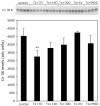
Figure 4. Cx 36 protein levels transiently decrease after SCI
Upper panel, immunoprecipitation followed by western blot of Cx 36 protein. Lower panel, quantification of the western blot (arbitrary units). Adult rats underwent spinal cord transection. After 7 (Tx + 7D), 14 (Tx + 14D), or 30 (Tx + 30D) days following transection, the entire lumbar region of the spinal cord was assayed for Cx 36 protein levels. 7 days following transection, one group of rats was treated with passive exercise for 30 days (Tx + Ex), while another group was given MOD for 30 days (Tx + MOD), and their Cx 36 protein levels assayed. There was a transient decrease in Cx 36 protein after 7 days, but it returned to normal levels over the course of a few weeks.

References

    1. Angel RW, Hofmann WW. The H Reflex in Normal, Spastic, and Rigid Subjects. Arch Neurol. 1963;9:591–596. - PubMed
    1. Arfaj A, Yates C, Reese NB, Ishida K, Skinner RD, Garcia-Rill E. Changes in the H-reflex after spinal cord injury: A longitudinal study in awake rats. Neurosci Abstr. 2007;33:405.22.
    1. Atsuta Y, Abraham P, Iwahara T, Garcia-Rill E, Skinner RD. Control of locomotion in vitro: II. Chemical stimulation. Somatosens Mot Res. 1991;8:55–63. - PubMed
    1. Atsuta Y, Garcia-Rill E, Skinner RD. Electrically induced locomotion in the in vitro brainstem-spinal cord preparation. Brain Res. 1988;470:309–312. - PubMed
    1. Ballon JS, Feifel D. A systematic review of modafinil: Potential clinical uses and mechanisms of action. J Clin Psychiatry. 2006;67:554–566. - PubMed

Publication types

LinkOut - more resources