. 2011 May;32(14):3564-74.
doi: 10.1016/j.biomaterials.2011.01.064. Epub 2011 Feb 21.

The performance of human mesenchymal stem cells encapsulated in cell-degradable polymer-peptide hydrogels

Affiliations

The performance of human mesenchymal stem cells encapsulated in cell-degradable polymer-peptide hydrogels

Sarah B Anderson et al. Biomaterials. 2011 May.

Abstract

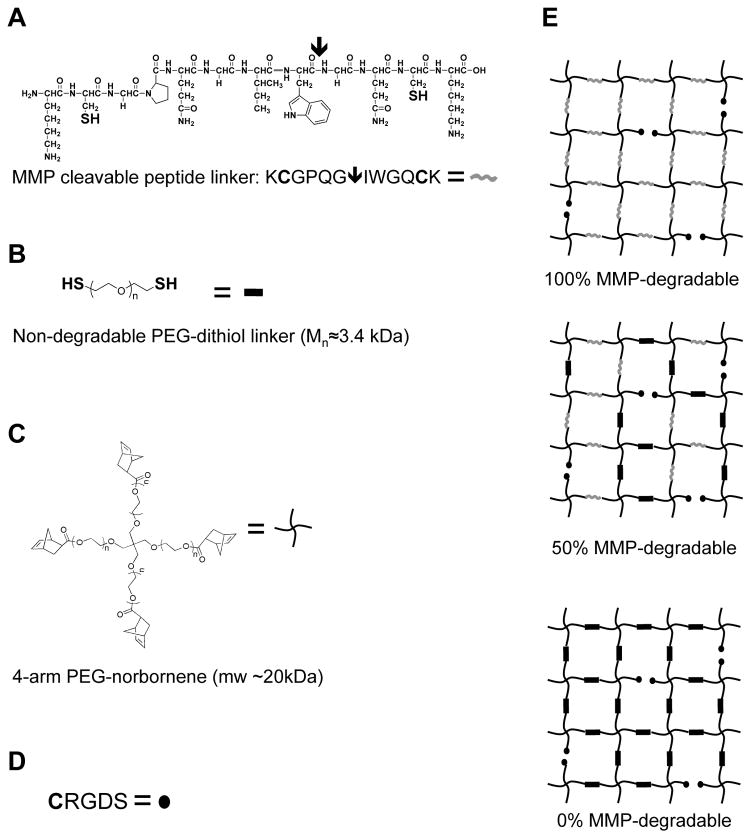
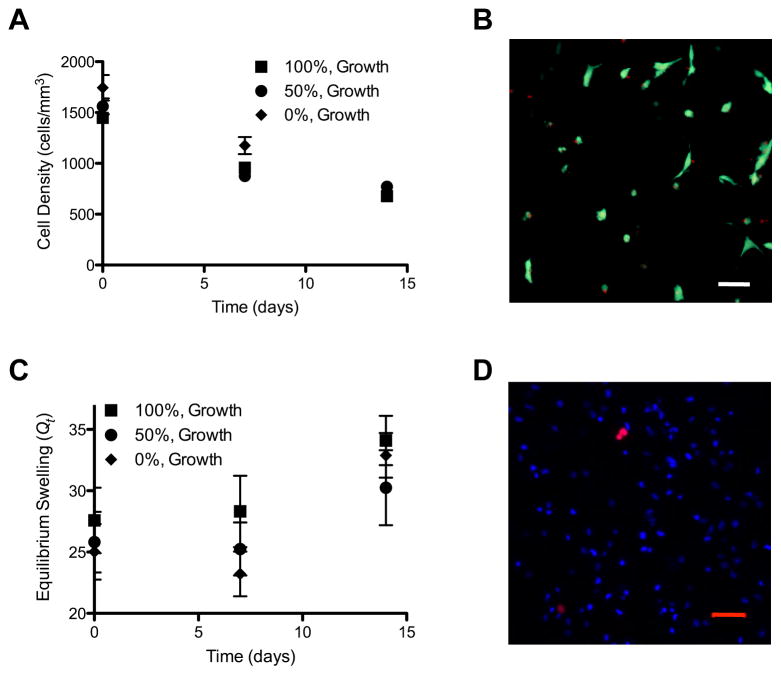
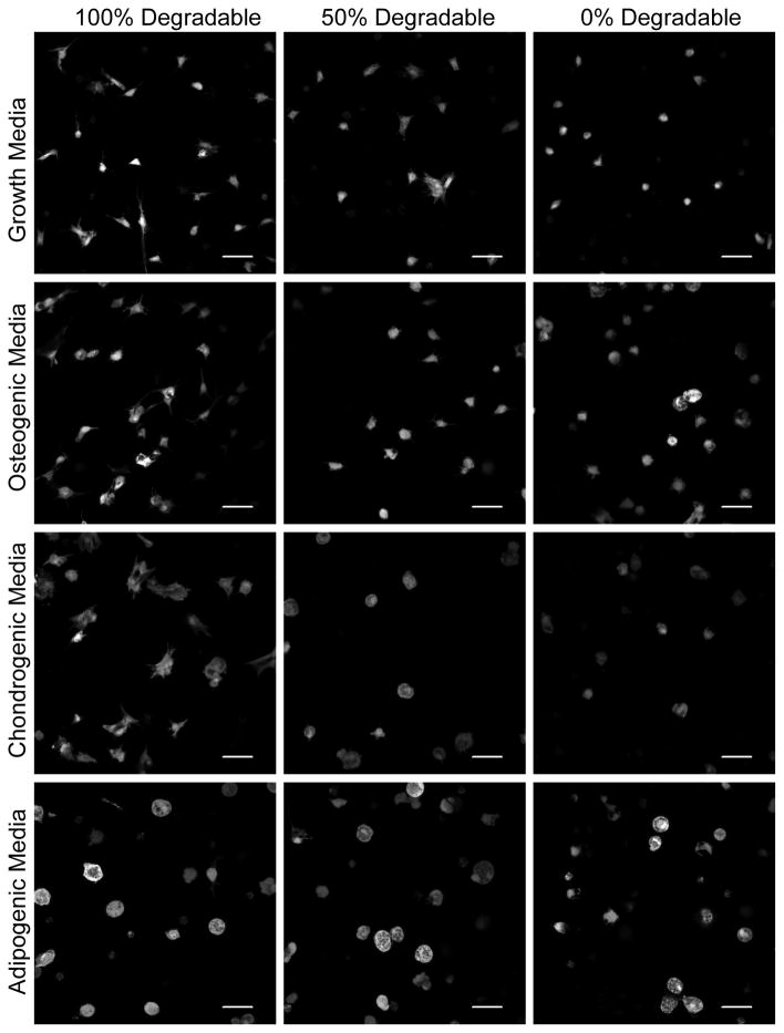
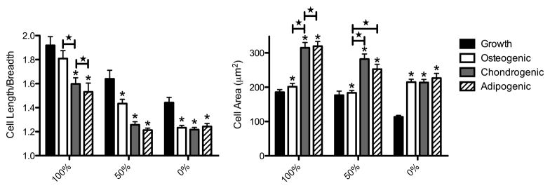
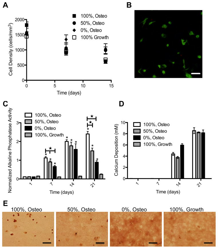
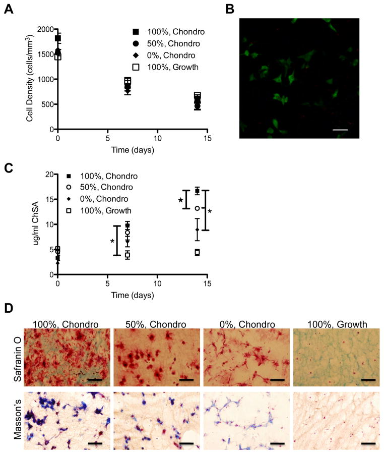
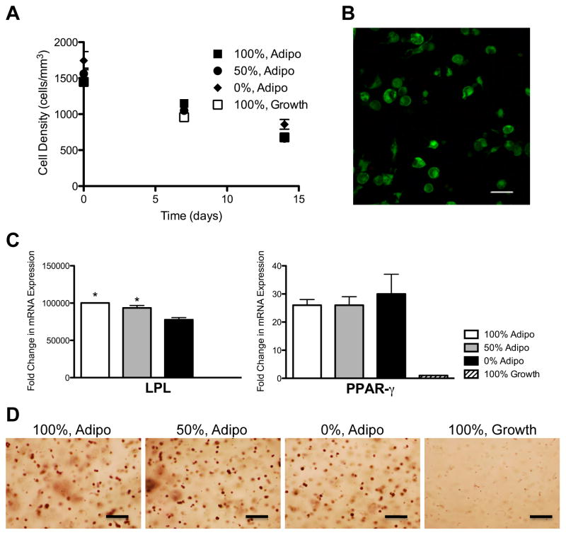
Thiol-ene photopolymerization offers a unique platform for the formation of peptide-functionalized poly(ethylene glycol) hydrogels and the encapsulation, culture and differentiation of cells. Specifically, this photoinitiated polymerization scheme occurs at neutral pH and can be controlled both spatially and temporally. Here, we have encapsulated human mesenchymal stem cells (hMSCs) in matrix metalloproteinase (MMP) degradable and cell-adhesive hydrogels using thiol-ene photopolymerization. We find that hMSCs survive equally well in this system, regardless of MMP-degradability. When hMSCs are encapsulated in these cell-degradable hydrogels, they survive and are able to proliferate. In classic hMSC differentiation medias, hMSCs locally remodel their microenvironment and take on characteristic morphologies; hMSCs cultured in growth or osteogenic differentiation media are less round, as measured by elliptical form factor, and are smaller than hMSCs cultured in chondrogenic or adipogenic differentiation media. In addition, hMSCs encapsulated in completely cell-degradable hydrogels and cultured in osteogenic, chondrogenic, or adipogenic differentiation media generally express increased levels of specific differentiation markers as compared to cells in hydrogels that are not cell-degradable. These studies demonstrate the ability to culture and differentiate hMSCs in MMP-degradable hydrogels polymerized via a thiol-ene reaction scheme and that increased cell-mediated hydrogel degradability facilitates directed differentiation of hMSCs.


Figures

Figure 1
Figure 1
Schematic of thiol-ene hydrogels with variable degradability
Figure 2
Figure 2
hMSCs encapsulated in MMP-degradable hydrogels survive and proliferate. hMSCs were encapsulated in MMP-degradable PEG-hydrogels, with varying degrees of degradability (percentages indicate extent of hydrogel linkers that are MMP-degradable). (A) Cell density was determined as the number of cells per mm3, at indicated times. (B) Representative image of hMSCs stained as live (Calcein AM/Green) or dead (EthD/Red) after 14 days of culture in growth media while encapsulated in 100% MMP-degradable PEG-hydrogels. (C) Equilibrium swelling ratio at indicated times (Qt). (D) Representative image of hMSCs encapsulated in 100% MMP-degradable PEG-hydrogels for 48 hours and labeled with Click-iT EdU cell proliferation marker (Red) and Hoescht DNA marker (Blue). Scale bars represent 50 μm.
Figure 3
Figure 3
Morphology of hMSCs encapsulated in MMP-degradable hydrogels. hMSCs encapsulated in MMP-degradable PEG-hydrogels, with varying degrees of degradability (percentages indicate extent of hydrogel linkers that are MMP-degradable), were stained with Calcein AM after 14 days of culture in indicated media and then imaged by confocal microscopy. Scale bar represents 50 μm.
Figure 4
Figure 4
hMSC circularity and spreading vary with hydrogel degradability and culture conditions. hMSCs encapsulated in MMP-degradable PEG-hydrogels, with varying degrees of degradability (percentages indicate extent of hydrogel linkers that are MMP-degradable), were stained with Calcein AM after 14 days of culture in indicated media and then imaged by confocal microscopy. Cell length divided by cell breadth was calculated to estimate circularity of cells, where 1.0 equals completely circular. Cell area was calculated to estimate the degree of spreading in hydrogels. * indicates significantly different from growth (p< 0.04). ★ indicates significantly different from each other (p<0.03). n>80.
Figure 5
Figure 5
hMSCs survive and express osteogenic markers in MMP-degradable hydrogels. hMSCs were encapsulated in PEG-hydrogels, with varying degrees of degradability (percentages indicate extent of hydrogel linkers that are MMP-degradable), and cultured in growth or osteogenic (Osteo) media. (A) Cell density was determined by staining encapsulated hMSCs with calcein AM and assaying cell density as the number of viable cells per mm3. (B) Representative image of hMSCs stained as live (Calcein AM/Green) or dead (EthD/Red) after 14 days of culture in osteogenic media while encapsulated in 100% MMP-degradable PEG-hydrogels. (C) Induction of alkaline phosphatase activity was normalized to alamar blue. * indicates significantly different from 100%, Growth condition (p<0.02). ★ indicates significantly different from each other (p<0.05). Deposition of mineralized matrix was assayed by (D) calcium chloride concentration and (E) alizarin red staining of fixed hydrogel cytrosections. Scale bars represent 50 μm.
Figure 6
Figure 6
hMSCs survive and express chondrogenic markers in MMP-degradable hydrogels. hMSCs were encapsulated in PEG-hydrogels, with varying degrees of degradability (percentages indicate extent of hydrogel linkers that are MMP-degradable), and cultured in growth or chondrogenic (Chondro) media. (A) Cell density was determined by staining encapsulated hMSCs with calcein AM and assaying cell density as measured by the number of viable cells per mm3. (B) Representative image of hMSCs stained as live (Calcein AM/Green) or dead (EthD/Red) after 14 days of culture in chondrogenic media while encapsulated in 100% MMP-degradable PEG-hydrogels. (C) Deposition of chondrogenic glycosaminoglycans was determined by DMMB assay as determined against a chondroitin sulfate standard. (D) Safranin O staining of hydrogel cryosections demonstrates GAGs in red, cytoplasm and background hydrogel staining in green and nuclie in black. Collagen deposition was demonstrated by Masson’s trichrome stain where collagen is stained blue, cytoplasm is stained red and nuclei are stained blue-black. * indicates significantly different from each other. ★ indicates significantly different from day 14 100%, Growth. (p<0.02). Scale bars represent 50 μm.
Figure 7
Figure 7
hMSCs survive and express adipogenic markers in MMP-degradable hydrogels. hMSCs were encapsulated in PEG-hydrogels, with varying degrees of degradability (percentages indicate extent of hydrogel linkers that are MMP-degradable), and cultured in growth or adipogenic (Adipo) media. (A) Cell density was determined by staining encapsulated hMSCs with calcein AM and assaying cell density as measured by the number of viable cells per mm3. (B) Representative image of hMSCs stained as live (Calcein AM/Green) or dead (EthD/Red) after 14 days of culture in growth media while encapsulated in 100% MMP-degradable PEG-hydrogels. (C) Expression of the adipogenic markers lipoprotein lipase (LPL) and peroxisome proliferator activated receptor gamma (PPAR-γ) were determined by qRT-PCR. * Indicates significantly different from 0%, Adipo (p<0.02). (D) Lipids were stained in fixed hydrogel cryosections using Oil Red O. Scale bars represent 50 μm.

References

    1. Tibbitt MW, Anseth KS. Hydrogels as extracellular matrix mimics for 3D cell culture. Biotechnol Bioeng. 2009;103:655–63. - PMC - PubMed
    1. Salinas CN, Anseth KS. The enhancement of chondrogenic differentiation of human mesenchymal stem cells by enzymatically regulated RGD functionalities. Biomaterials. 2008;29:2370–7. - PMC - PubMed
    1. Salinas CN, Anseth KS. Decorin moieties tethered into PEG networks induce chondrogenesis of human mesenchymal stem cells. J Biomed Mater Res A. 2009;90:456–64. - PMC - PubMed
    1. Lutolf MP, Hubbell JA. Synthesis and physicochemical characterization of end-linked poly(ethylene glycol)-co-peptide hydrogels formed by michael-type addition. Biomacromolecules. 2003;4:713–22. - PubMed
    1. Fairbanks BD, Schwartz MP, Halevi AE, Nuttelman CR, Bowman CN, Anseth KS. A versatile synthetic extracellular matrix mimic via thiol-norbornene photopolymerization. Adv Mat. 2009;21:5005–10. - PMC - PubMed

Publication types