. 2011 Jun;1808(6):1701-8.
doi: 10.1016/j.bbamem.2011.02.009. Epub 2011 Feb 18.

Disrupting microtubule network immobilizes amoeboid chemotactic receptor in the plasma membrane

Affiliations

Disrupting microtubule network immobilizes amoeboid chemotactic receptor in the plasma membrane

S de Keijzer et al. Biochim Biophys Acta. 2011 Jun.

Abstract

Signaling cascades are initiated in the plasma membrane via activation of one molecule by another. The interaction depends on the mutual availability of the molecules to each other and this is determined by their localization and lateral diffusion in the cell membrane. The cytoskeleton plays a very important role in this process by enhancing or restricting the possibility of the signaling partners to meet in the plasma membrane. In this study we explored the mode of diffusion of the cAMP receptor, cAR1, in the plasma membrane of Dictyostelium discoideum cells and how this is regulated by the cytoskeleton. Single-particle tracking of fluorescently labeled cAR1 using Total Internal Reflection Microscopy showed that 70% of the cAR1 molecules were mobile. These receptors showed directed motion and we demonstrate that this is not because of tracking along the actin cytoskeleton. Instead, destabilization of the microtubules abolished cAR1 mobility in the plasma membrane and this was confirmed by Fluorescence Recovery after Photobleaching. As a result of microtubule stabilization, one of the first downstream signaling events, the jump of the PH domain of CRAC, was decreased. These results suggest a role for microtubules in cAR1 dynamics and in the ability of cAR1 molecules to interact with their signaling partners.


Figures

Figure 1
Figure 1. Analysis of cAR1 mobility
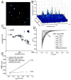
(A) Image from a stack (movie M1, see supplemental material) showing signals form single cAR1-Halo-TMR molecules in the basal membrane of live Dictyostelium cells as imaged with Total Internal Reflection Fluorescence Microscopy (n=19 cells). (B) The 3D graph of the intensities of the single molecules. (C) Representive trajectories of two different dynamic behaviors, mobile (1) and immobile (2), of cAR1. (D) Cumulative probability distributions (P(r2)) of the square displacements (r2) of cAR1-Halo-TMR at different tlag. The PSD were described with a biexponential fit (dashed curve) giving two Mean Square Displacements (ri2(t)) and the fraction size (α) at each tlag. (E) MSD data of single cAR1 molecules versus tlag. The cAR1-Halo-TMR data for the immobile fraction (30% of the molecules) was close to the error in positional accuracy (light grey dashed line) 4σ2= 0.0025 μm2. The mobile fraction (n=59 trajectories) was described by a model describing diffusion with flow (grey dashed line). The fitting results were D=0.015 ± 0.002μm2/s and velocity of v= 0.16 ± 0.02 μm/s.
Figure 2
Figure 2. Disruption of actin cytoskeleton increases cAR1 mobility
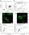
(A) Trajectory of a cAR1 molecule after 10 min incubation with 5 μM Latrunculin (n=20 cells). (B) After Latrunculin treatment there were two populations of cAR1 molecules and the fraction sizes were not changed, 30% immobile and 70% mobile. The mobile population still showed directed movement, but the speed of movement was increased D=0.017 ± 0.002μm2/s and a velocity of v= 0.28 ± 0.03 μm/s (n=21 trajectories)
Figure 3
Figure 3. Destabilization of microtubules abolishes cAR1 movement
(A) Trajectory of a cAR1 molecule after 10 min incubation with 50 μM Benomyl (n=19 cells), the insert shows the trajectory on a smaller (10x) scale. (B) After destabilization of the microtubules with Benomyl there was only one population of cAR1-Halo-TMR molecules and when compared to the slow population of cAR1-Halo-TMR molecules (black squares), these were all completely immobile after addition of Benomyl (black circles). (C) Microtubules were visualized with tubulin-GFP. (D) After 10 min of incubation with 50 mM Benomyl the aster-like assemblies of microtubules were no longer observed. The microtubules were decomposed and a homogeneous distribution of tubulin-GFP could be seen in the cells. Fluorescence recovery curves of the lipid dye FM4-64 (E) and cAR1-eYFP (F) before (black) and after Benomyl addition (red) (n>10 cells for each condition). The recovery of the fluorescence after photobleaching within a circle of 1μm diameter of the basal plasma membrane is plotted against time. Different time intervals were used for FM4-64 and cAR1-eYFP.
Figure 4
Figure 4. Downstream signaling disrupted with benomyl
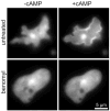
Translocation of PH-GFP to the plasma membrane in the absence (A) or presence of Benomyl (B). Upon cAMP addition there is a jump of PH-GFP (from CRAC) to the plasma membrane in 35 of the observed 51 cells (n=4 experiments). In the presence of Benomyl there was no PH-jump in the 41 observed cells. Images were from movies (see supplemental movies M2 and M3)

Similar articles

Cited by

References

    1. Pierce KL, Premont RT, Lefkowitz RJ. Seven-transmembrane receptors. Nat Rev Mol Cell Biol. 2002;3:639–650. - PubMed
    1. Hamm HE. How activated receptors couple to G proteins. Proc Natl Acad Sci U S A. 2001;98:4819–4821. - PMC - PubMed
    1. Sun Y, McGarrigle D, Huang XY. When a G protein-coupled receptor does not couple to a G protein. Mol Biosyst. 2007;3:849–854. - PubMed
    1. Milne JL, Wu L, Caterina MJ, Devreotes PN. Seven helix cAMP receptors stimulate Ca2+ entry in the absence of functional G proteins in Dictyostelium. J Biol Chem. 1995;270:5926–5931. - PubMed
    1. Hall RA, Premont RT, Lefkowitz RJ. Heptahelical receptor signaling: beyond the G protein paradigm. J Cell Biol. 1999;145:927–932. - PMC - PubMed

Publication types

MeSH terms