Histotripsy fractionation of prostate tissue: local effects and systemic response in a canine model
- PMID: 21334667
- PMCID: PMC3075837
- DOI: 10.1016/j.juro.2010.11.044
Histotripsy fractionation of prostate tissue: local effects and systemic response in a canine model
Abstract
Purpose: Histotripsy is an extracorporeal ultrasound technology that uses cavitational mechanisms to produce nonthermal tissue destruction. Previously we reported the feasibility of histotripsy for prostate tissue fractionation and immediate debulking. In this study we characterized the local effects and systemic response after histotripsy treatment of prostate tissue in an in vivo canine model.
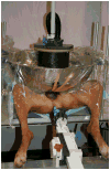
Materials and methods: Histotripsy was applied transabdominally to the prostate in 18 intact male canine subjects under general anesthesia. Acoustic bursts (4 μseconds) were delivered at a 300 Hz pulse repetition rate from a highly focused 750 kHz piezoelectric ultrasound transducer with a 15 cm aperture and 3 × 3 × 8 mm focal volume. Specimens of the prostate and surrounding structures were obtained at prescribed time points (0, 7, 28 or 56 days) after histotripsy. Blood and urine parameters were assessed periodically while clinical evaluation incorporating a validated veterinary pain scale was performed daily.
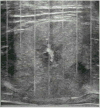
Results: Conventional transrectal ultrasound facilitated targeting of the focal volume and provided real-time assessment of cavitation activity. Fractionation of the targeted volume and clearance of the resultant debris with urination produced a treatment cavity in each prostate. No acoustic collateral damage was seen and urothelialization of the treatment cavity developed within 28 days of treatment. Only transient laboratory value abnormalities and minimal hematuria were noted after treatment. Pain scores revealed only mild posttreatment discomfort.
Conclusions: Histotripsy produced consistent tissue fractionation and prostate debulking without collateral acoustic injury or clinical side effects and it was well tolerated in the canine model.
Copyright © 2011 American Urological Association Education and Research, Inc. Published by Elsevier Inc. All rights reserved.
Figures


Similar articles
-
Histotripsy of the prostate using a commercial system in a canine model.J Urol. 2014 Mar;191(3):860-5. doi: 10.1016/j.juro.2013.08.077. Epub 2013 Sep 6. J Urol. 2014. PMID: 24012583
-
Histotripsy: minimally invasive technology for prostatic tissue ablation in an in vivo canine model.Urology. 2008 Sep;72(3):682-6. doi: 10.1016/j.urology.2008.01.037. Epub 2008 Mar 17. Urology. 2008. PMID: 18342918 Free PMC article.
-
Histotripsy of the prostate: dose effects in a chronic canine model.Urology. 2009 Oct;74(4):932-7. doi: 10.1016/j.urology.2009.03.049. Epub 2009 Jul 22. Urology. 2009. PMID: 19628261 Free PMC article.
-
Development and translation of histotripsy: current status and future directions.Curr Opin Urol. 2014 Jan;24(1):104-10. doi: 10.1097/MOU.0000000000000001. Curr Opin Urol. 2014. PMID: 24231530 Free PMC article. Review.
-
Advances in Tumor Management: Harnessing the Potential of Histotripsy.Radiol Imaging Cancer. 2024 May;6(3):e230159. doi: 10.1148/rycan.230159. Radiol Imaging Cancer. 2024. PMID: 38639585 Free PMC article. Review.
Cited by
-
Robotically Assisted Sonic Therapy (RAST) for Noninvasive Hepatic Ablation in a Porcine Model: Mitigation of Body Wall Damage with a Modified Pulse Sequence.Cardiovasc Intervent Radiol. 2019 Jul;42(7):1016-1023. doi: 10.1007/s00270-019-02215-8. Epub 2019 Apr 30. Cardiovasc Intervent Radiol. 2019. PMID: 31041527 Free PMC article.
-
Urethral-sparing histotripsy of the prostate in a canine model.Urology. 2012 Sep;80(3):730-5. doi: 10.1016/j.urology.2012.05.027. Epub 2012 Jul 26. Urology. 2012. PMID: 22840869 Free PMC article.
-
Effects of Thermal Preconditioning on Tissue Susceptibility to Histotripsy.Ultrasound Med Biol. 2015 Nov;41(11):2938-54. doi: 10.1016/j.ultrasmedbio.2015.07.016. Epub 2015 Aug 28. Ultrasound Med Biol. 2015. PMID: 26318560 Free PMC article.
-
Focused ultrasound: tumour ablation and its potential to enhance immunological therapy to cancer.Br J Radiol. 2018 Feb;91(1083):20170641. doi: 10.1259/bjr.20170641. Epub 2018 Jan 17. Br J Radiol. 2018. PMID: 29168922 Free PMC article. Review.
-
Immunological Effects of Histotripsy for Cancer Therapy.Front Oncol. 2021 May 31;11:681629. doi: 10.3389/fonc.2021.681629. eCollection 2021. Front Oncol. 2021. PMID: 34136405 Free PMC article. Review.
References
-
- Ahyai SA, Lehrich K, Kuntz RM. Holmium laser enucleation versus transurethral resection of the prostate: 3-year follow-up results of a randomized clinical trial. Eur Urol. 2007;52:1456. - PubMed
-
- Spaliviero M, Araki M, Wong C. Short term outcomes of greenlight HPS laser photoselective vaporization prostatectomy (PVP) for benign prostatic hyperplasia (BPH) J Endourol. 2008;10:2341. - PubMed
-
- Mebust WK, Holtgrewe HL, Cockett AT, et al. Transurethral prostatectomy: immediate and postoperative complications: A cooperative study of 13 participating institutions evaluating 3,885 patients. J Urol. 1989;141:243. - PubMed
-
- Roos NP, Wennberg JE, Malenka DJ, et al. Mortality and reoperation after open and transurethral resection of the prostate for benign prostatic hyperplasia. N Engl J Med. 1989;320:1120. - PubMed
-
- Bouchier-Hayes DM, Van Appledorn S, Bugeja P, et al. A randomized trial of photoselective vaporization of the prostate using the 80-W potassium-titanyl-phosphate laser vs transurethral prostatectomy, with a 1-year follow-up. BJU International. 2009;105:964–9. - PubMed
Publication types
MeSH terms
Grants and funding
LinkOut - more resources
Full Text Sources
Other Literature Sources
Medical

