. 2011 Feb 18;144(4):577-89.
doi: 10.1016/j.cell.2011.01.014.

The basement membrane of hair follicle stem cells is a muscle cell niche

Affiliations

The basement membrane of hair follicle stem cells is a muscle cell niche

Hironobu Fujiwara et al. Cell. .

Abstract

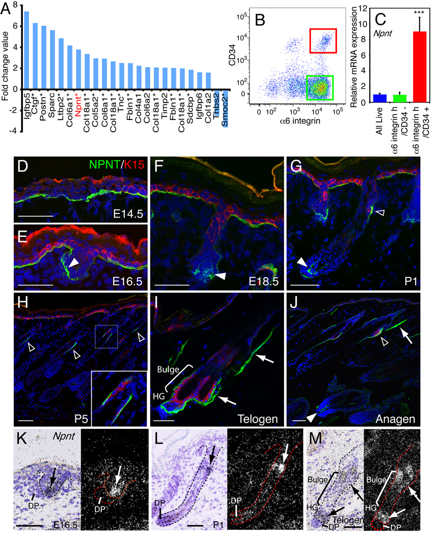
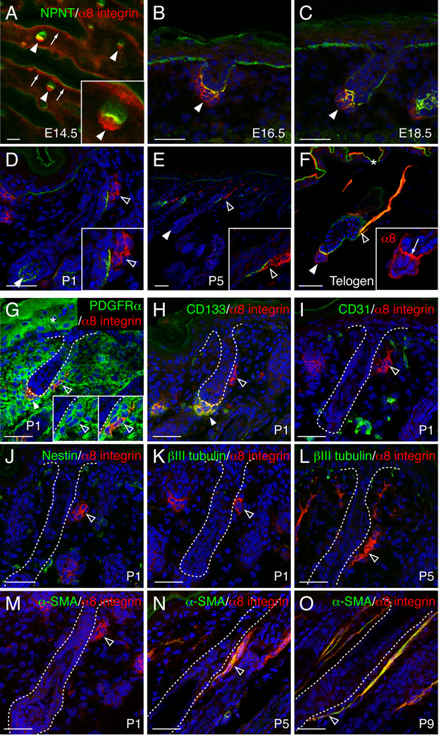
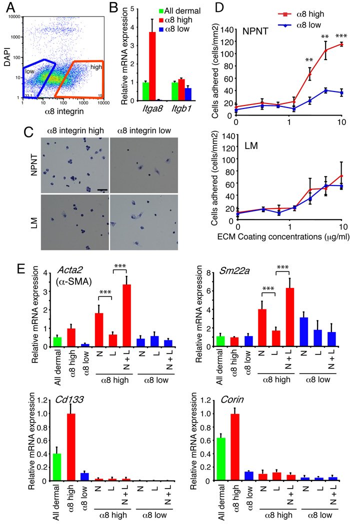
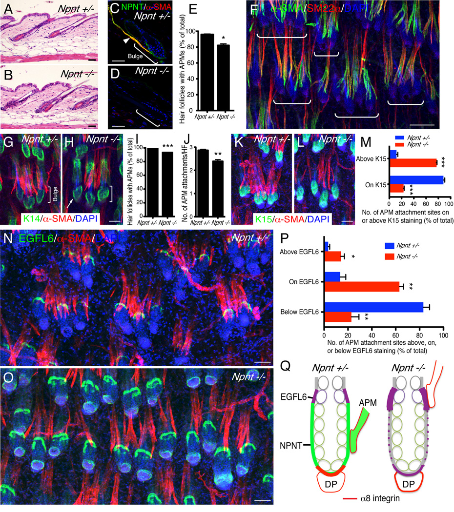
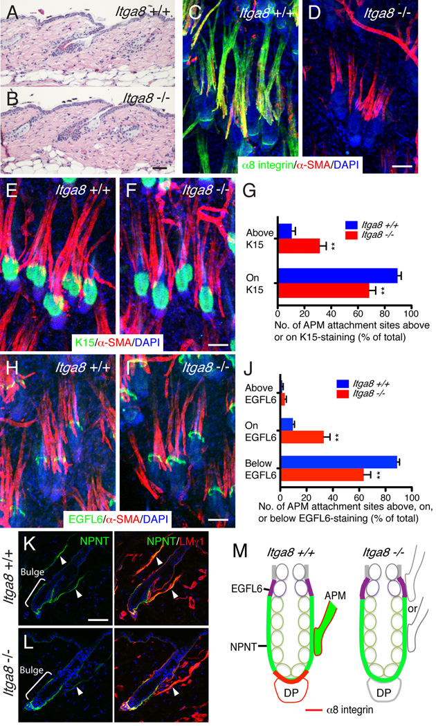
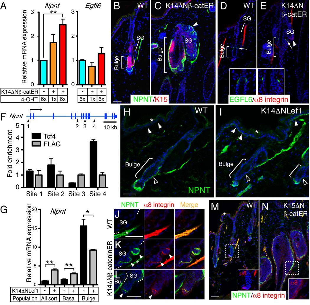
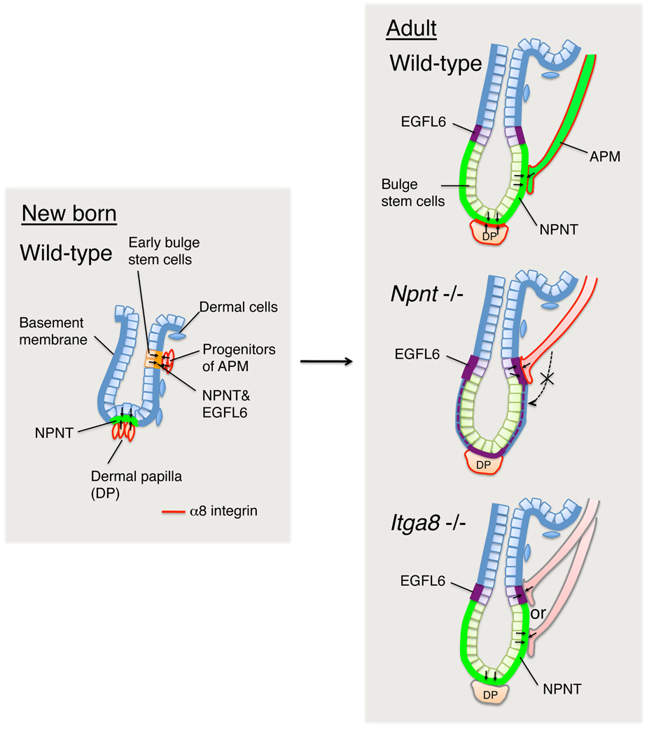
The hair follicle bulge in the epidermis associates with the arrector pili muscle (APM) that is responsible for piloerection ("goosebumps"). We show that stem cells in the bulge deposit nephronectin into the underlying basement membrane, thus regulating the adhesion of mesenchymal cells expressing the nephronectin receptor, α8β1 integrin, to the bulge. Nephronectin induces α8 integrin-positive mesenchymal cells to upregulate smooth muscle markers. In nephronectin knockout mice, fewer arrector pili muscles form in the skin, and they attach to the follicle above the bulge, where there is compensatory upregulation of the nephronectin family member EGFL6. Deletion of α8 integrin also abolishes selective APM anchorage to the bulge. Nephronectin is a Wnt target; epidermal β-catenin activation upregulates epidermal nephronectin and dermal α8 integrin expression. Thus, bulge stem cells, via nephronectin expression, create a smooth muscle cell niche and act as tendon cells for the APM. Our results reveal a functional role for basement membrane heterogeneity in tissue patterning. PAPERCLIP:


Figures

Figure 1
Figure 1. Nephronectin expression in skin
(A) ECM genes that are upregulated or downregulated in bulge stem cells relative to other basal keratinocytes, ranked based on log2 fold change value (see Table S1). Asterisks indicate the genes that are also upregulated or downregulated in mouse label retaining cells (Tables S2, S3). Some genes are listed more than once due to their multiple spots on the array. (B, C) Adult telogen dorsal keratinocytes were FACS-sorted according to α6 integrin and CD34 expression (B). Bulge stem cells (red gate; α6 integrinhigh/CD34+), non-bulge basal stem cells (green gate; α6 integrinhigh/CD34−) and all live basal cells were sorted and Npnt mRNA levels were determined by Q-PCR (C). Data are mean ± SEM from three mice. (D–J) Sections of E14.5 (D), E16.5 (E), E18.5 (F), P1 (G), P5 (H), adult telogen (I) and anagen (J) skin were immunostained for nephronectin (NPNT; green) and bulge stem cell marker K15 (red), with DAPI counterstain (blue). Note nephronectin deposition in hair germ (white arrowheads), bulge (open arrowheads) and APM (arrows). (K–M) In situ hybridization with Npnt probe on E16.5 (K), P1 (L), and adult telogen skin (M). Arrows indicate the strong nephronectin expression. Scale bars: 50 µm. See also Figure S1.
Figure 2
Figure 2. Nephronectin colocalizes with α8 integrin-positive dermal cells in dermal papilla and arrector pili muscle
(A–F) Immunostaining for nephronectin (green) and α8 integrin (red) in developing and adult dorsal skin. Arrows: α8 expression in superficial dermis (A) or at nephronectin-positive basement membrane between hair germ and dermal papilla (F); white arrowheads: dermal papilla cells; open arrowheads: α8 integrin-positive cells around bulge. (G–O) Expression of α8 integrin (red) and other dermal markers (green) in postnatal dorsal skin. Open arrowheads: α8 integrin-positive dermal cells adjacent to bulge. White arrowhead in G and H: dermal papilla. All sections were counterstained with DAPI (blue). Asterisk indicates non-specific staining. Scale bars: 50 µm. See also Figure S2.
Figure 3
Figure 3. Adhesion and gene expression of dermal cells plated on nephronectin
(A, B) Disaggregated P1 dermal cells were FACS-sorted on the basis of α8 integrin levels (A), confirmed by Q-PCR for α8 integrin (Itga8) and β1 integrin (Itgb1) (B). Data are means ± SEM from three mice. (C, D) Solid-phase cell adhesion assays with FACS-sorted dermal cells plated on nephronectin (NPNT)- or laminin (LM)-coated dishes (10 µg/ml). Cells were stained with Diff Quik (C) and quantitated (D). (C) Scale bar: 50 µm. (D) Data are means ± SEM from triplicate wells. (E) Q-PCR of smooth muscle (Acta2 and Sm22a) and dermal papilla marker (Cd133 and Corin) expression in unfractionated (all dermal), α8 integrin-high or -low populations seeded on ECM protein-coated dishes (10 µg/ml of each protein) for 12 hours. N: nephronectin; L: laminin; N+L: nephronectin and laminin. Data are means ± SEM from three independent wells.
Figure 4
Figure 4. Nephronectin is required to anchor arrector pili muscles to the bulge
(A, B) H&E-stained adult dorsal telogen skin. (C–E) Adult dorsal skin immunostained for nephronectin (green) and α-SMA (red) and counterstained with DAPI (blue). Arrowhead indicates APM. (E) % hair follicles with arrector pili muscles. (F) Maximum projection image of dorsal wild type skin whole-mount stained for α-SMA (green), SM22α (red), and nuclei (blue). White brackets indicate groups of hair follicles that share arrector pili muscles. (G–J) Whole-mounts (G, H) were immunostained for K14 (green) and α-SMA (red), with DAPI counterstain (blue). Arrow (H) indicates hair follicle without associated APM. Brackets indicate bulge. % follicles with arrector pili muscles (I) and number of APM attachments per hair follicle (J). (K–M) Whole-mounts were immunostained for K15 (green) and α-SMA (red), with DAPI counterstain (blue). Position of APM attachment sites relative to K15-positive bulge was quantified (M). (N–P) Whole-mounts were immunostained for EGFL6 (green) and α-SMA (red), with DAPI counterstain (blue). Position of APM attachment sites relative to EGFL6-positive zone was quantified (P). (Q) Schematic summary of data. Green: nephronectin; purple: EGFL6; red: α8 integrin; green circles: K15-positive bulge cells. In the presence of nephronectin, arrector pili muscles insert at the bulge. In the absence of nephronectin, EGFL6 expression in the upper bulge is increased and muscles insert in that region. α8 integrin colocalises with nephronectin in the basement membrane of the hair germ adjacent to the dermal papilla (DP). In the absence of nephronectin EGFL6 is expressed in that region, but there is no colocalisation with α8 integrin. All skin samples were from the back of 7 week old telogen mice. (E, I, J, M, P) Data are means ± SEM from three mice, 100 follicles per mouse. Scale bars: 50 µm; except for F (100 µm). See also Figure S3.
Figure 5
Figure 5. Deletion of α8 integrin disrupts specificity of APM attachment site to the bulge
(A, B) H&E-stained adult dorsal telogen skin. (C, D) Skin whole-mounts were immunostained for α8 integrin (green) and α-SMA (red), with DAPI counterstain (blue). (E–G) Whole-mounts (E, F) were immunostained for K15 (green) and α-SMA (red), with DAPI counterstain (blue). Position of APM attachment sites relative to K15-positive bulge was quantified (G). (H–J) Whole-mounts were immunostained for EGFL6 (green) and α-SMA (red), with DAPI counterstain (blue). Position of APM attachment sites relative to EGFL6-positive zone was quantified (J). (K) Conventional cryo-sections were immunostained for nephronectin (green) and laminin γ1 chain (red), with DAPI counterstain (blue). Arrowheads indicate APM. (M) Schematic summary of data. Green: nephronectin; purple: EGFL6; red: α8 integrin; green circles: K15-positive bulge cells. In the presence of α8 integrin, the APM is anchored to the bulge. In the absence of α8 integrin, muscles lose specificity for nephronectin and can anchor to both the nephronectin-positive bulge and the EGFL6-positive upper bulge regions. In the absence of α8 integrin, nephronectin deposition in the APM is disrupted, but that in the bulge is unaffected. All skin samples were from the back of 7–11 week old telogen mice. (G, J) Data are means ± SEM from four mice, 100 follicles per mouse. Scale bars: 50 µm. See also Figure S4.
Figure 6
Figure 6. Regulation of regional nephronectin-α8 integrin interaction by Wnt/β-catenin signalling
(A) Q-PCR of Npnt and Egfl6 mRNA in keratinocytes isolated from skin of wild type (ΔNβ-catER −) and K14ΔNβ-cateninER mice (ΔNβ-catER +) that had been treated with 4-OHT for the number of times indicated. Data are means ± SEM from three mice. (B–E) Dorsal skin of wild type and K14ΔNβ-cateninER mice treated 6× with 4-OHT and immunostained for nephronectin (green; B, C) or EGFL6 (green; D, E) and K15 (red; B, C) or α8 integrin (red; D, E), with DAPI counterstain (blue). Arrowheads indicate ectopic hair follicle arising from sebaceous gland (SG). Arrows indicate EGFL6- staining. Inserts show higher magnification views of EGFL6 staining around the bulge. (F) ChIP using antibodies against Tcf4 and FLAG in K14ΔNβ-cateninER keratinocytes. Primers surrounding four conserved putative binding sites for Lef/Tcfs at the Npnt locus were used to detect the precipitated DNA fragments. Data are means ± SEM of two independent experiments. (G) Q-PCR of mRNA from FACS-isolated bulge, non-bulge (basal) and total basal (All sort) keratinocytes from wild type and K14ΔNLef1 adult telogen skin. Data are means ± SEM from three mice. (H, I) Wild type and K14ΔNLef1 skin immunostained for nephronectin (green) with DAPI counterstain (blue). Basement membrane of interfollicular epidermis (white arrowheads) and bulge (open arrowheads) is indicated. (J–N) Sections of wild type (J, M), K14ΔNβ-cateninER (K, L) and K15ΔNβ-cateninER (N) skin treated 6× (J–L) or 9× (M, N) with 4-OHT and immunostained for nephronectin (green) and α8 integrin (red), with DAPI counterstain (blue). Arrowheads indicate ectopic hair follicles. Asterisks in J and K indicate arrector pili muscles. Inserts in M, N show magnified images of α8 integrin staining around the bulge. Asterisks in H, I and M indicate non-specific staining. Scale bars: 50 µm. See also Figure S5.
Figure 7
Figure 7. Model depicting role for nephronectin-α8β1 interactions in creating the niche for the arrector pili muscle at the hair follicle bulge
During hair morphogenesis in neonatal skin, early bulge stem cells locally deposit nephronectin in the bulge basement membrane. Nephronectin induces neighbouring mesenchymal progenitors to differentiate into α8 integrin-positive APM cells, which adhere specifically to nephronectin, establishing a stable anchorage to the bulge that is maintained throughout adult life. In the absence of nephronectin, the APM is not anchored to the bulge, but attaches above the bulge, where there is compensatory upregulation of EGFL6. Lack of nephronectin also disturbs α8 integrin-mediated hair follicle-dermal papilla interactions (Figure S3). In the absence of α8 integrin, nephronectin is still deposited in the bulge, but the selectivity of the APM interaction is lost and muscles are anchored both to the nephronectin-positive bulge and the EGFL6-positive upper bulge.

References

    1. Akiyama M, Dale BA, Sun TT, Holbrook KA. Characterization of hair follicle bulge in human fetal skin: the human fetal bulge is a pool of undifferentiated keratinocytes. J Invest Dermatol. 1995;105:844–850. - PubMed
    1. Alonso L, Fuchs E. Stem cells in the skin: waste not, Wnt not. Genes Dev. 2003;17:1189–1200. - PubMed
    1. Baker CM, Verstuyf A, Jensen KB, Watt FM. Differential sensitivity of epidermal cell subpopulations to beta-catenin-induced ectopic hair follicle formation. Dev Biol. 2010;343:40–50. - PMC - PubMed
    1. Barcaui CB, Pineiro-Maceira J, de Avelar Alchorne MM. Arrector pili muscle: evidence of proximal attachment variant in terminal follicles of the scalp. Br J Dermatol. 2002;146:657–658. - PubMed
    1. Bolcato-Bellemin AL, Lefebvre O, Arnold C, Sorokin L, Miner JH, Kedinger M, Simon-Assmann P. Laminin alpha5 chain is required for intestinal smooth muscle development. Dev Biol. 2003;260:376–390. - PubMed

Publication types