

doi: 10.1128/AEM.00286-11.
Epub 2011 Feb 18.
Characterization of lateral flagella of Selenomonas ruminantium
Affiliations
- PMID: 21335384
- PMCID: PMC3126368
- DOI: 10.1128/AEM.00286-11
Item in Clipboard
Characterization of lateral flagella of Selenomonas ruminantium
Appl Environ Microbiol.
2011 Apr.
Abstract
Selenomonas ruminantium produces a tuft of flagella near the midpoint of the cell body and swims by rotating the cell body along the cell's long axis. The flagellum is composed of a single kind of flagellin, which is heavily glycosylated. The hook length of S. ruminantium is almost double that of Salmonella.
Figures
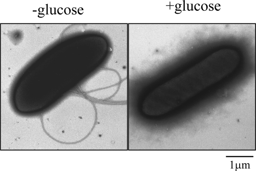
Electron microscopic images of Selenomonas ruminantium. Cells were grown in the absence of glucose (−glucose, left) or in the presence of glucose (+glucose, right).
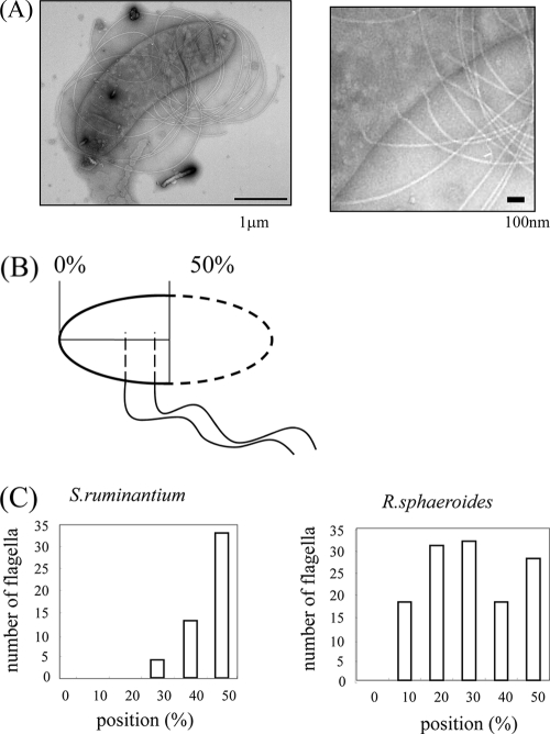
Distribution of flagella on a cell. (A) Electron microscopic image of a cell that was osmotically shocked and stained with 1% phosphotungstic acid (PTA). The flagellar form is coiled (left). Enlarged image of the left panel showing flagellar basal bodies in the membranes (right). (B) Schematic presentation showing how the positions of flagella on a cell were measured. (C) Diagram of the distributions of sites of flagellar protrusion on a cell for S. ruminantium (left) and R. sphaeroides (right). The total numbers of flagella counted were 50 and 127, respectively.
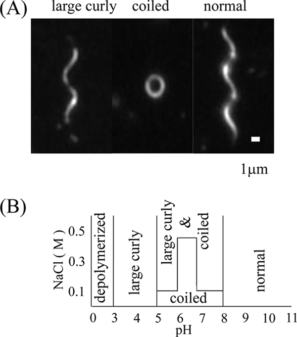
Flagellar polymorphic transition. (A) Dark-field microscopic images of polymorphs observed under various pH and salt conditions. The upper side of the helix was focused to show the handedness. (B) Schematic phase diagram of polymorphs observed at different pHs and NaCl concentrations.
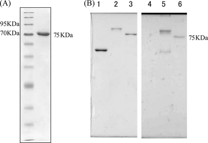
The S. ruminantium flagellin is glycosylated. (A) SDS gel pattern of an S. ruminantium flagellin. First lane, molecular size marker; second lane, single band of flagellin. (B) Detection of glycosylation of an S. ruminantium flagellin (lanes 3 and 6). Salmonella SJW1103 flagellin was used as a negative control (lanes 1 and 4), and Azospirillum flagellin was used as a positive control (lanes 2 and 5). Coomassie brilliant blue (CBB) staining (left) and PAS staining (right) of the purified flagellins are shown.
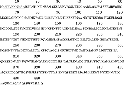
Amino acid sequence deduced from the S. ruminantium fliC1 gene. The underlined sequences were obtained by an analysis of purified FliC.
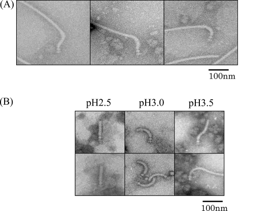
Shape and length of the hook. (A) Electron microscopic images of the hook basal body isolated from S. ruminantium. The desalted samples were stained with 2% PTA at pH 7.0. (B) Polymorphs of the hook under different pH conditions.
Similar articles
-
Structure of the Na+-driven flagellum from the homoacetogenic bacterium Acetobacterium woodii.FEBS Lett. 1998 Sep 4;434(3):325-8. doi: 10.1016/s0014-5793(98)01008-4. FEBS Lett. 1998. PMID: 9742948
-
Total reconstitution of Salmonella flagellar filaments from hook and purified flagellin and hook-associated proteins in vitro.J Mol Biol. 1989 Sep 5;209(1):109-14. doi: 10.1016/0022-2836(89)90174-5. J Mol Biol. 1989. PMID: 2810363
-
Flagellar growth in a filament-less Salmonella fliD mutant supplemented with purified hook-associated protein 2.J Biochem. 1993 Jul;114(1):39-44. doi: 10.1093/oxfordjournals.jbchem.a124136. J Biochem. 1993. PMID: 8407873
-
The archaeal flagellum: a different kind of prokaryotic motility structure.FEMS Microbiol Rev. 2001 Apr;25(2):147-74. doi: 10.1111/j.1574-6976.2001.tb00575.x. FEMS Microbiol Rev. 2001. PMID: 11250034 Review.
-
Bacterial and archaeal flagella as prokaryotic motility organelles.Biochemistry (Mosc). 2004 Nov;69(11):1203-12. doi: 10.1007/s10541-005-0065-8. Biochemistry (Mosc). 2004. PMID: 15627373 Review.
Cited by
-
Application of propionate-producing bacterial consortium in ruminal methanogenesis inhibited environment with bromoethanesulfonate as a methanogen direct inhibitor.Front Vet Sci. 2024 Oct 9;11:1422474. doi: 10.3389/fvets.2024.1422474. eCollection 2024. Front Vet Sci. 2024. PMID: 39444738 Free PMC article.
-
YSMR: a video tracking and analysis program for bacterial motility.BMC Bioinformatics. 2020 Apr 29;21(1):166. doi: 10.1186/s12859-020-3495-9. BMC Bioinformatics. 2020. PMID: 32349658 Free PMC article.
-
Bacterial motility: machinery and mechanisms.Nat Rev Microbiol. 2022 Mar;20(3):161-173. doi: 10.1038/s41579-021-00626-4. Epub 2021 Sep 21. Nat Rev Microbiol. 2022. PMID: 34548639 Review.
-
Novel methods for analysing bacterial tracks reveal persistence in Rhodobacter sphaeroides.PLoS Comput Biol. 2013 Oct;9(10):e1003276. doi: 10.1371/journal.pcbi.1003276. Epub 2013 Oct 24. PLoS Comput Biol. 2013. PMID: 24204227 Free PMC article.
-
The polar and lateral flagella from Plesiomonas shigelloides are glycosylated with legionaminic acid.Front Microbiol. 2015 Jun 26;6:649. doi: 10.3389/fmicb.2015.00649. eCollection 2015. Front Microbiol. 2015. PMID: 26167161 Free PMC article.
References
-
- Adler J., Templeton B. 1967. The effect of environmental conditions on the motility of Escherichia coli. J. Gen. Microbiol. 46:175–184 - PubMed
-
- Aizawa S.-I. 2009. Flagella, p. 393–403 In Schaechter M. (ed.), Encyclopedia of microbiology. Elsevier, Oxford, United Kingdom
MeSH terms
Substances
LinkOut - more resources
Full Text Sources
