Review
. 2011 Feb 18;108(4):512-26.
doi: 10.1161/CIRCRESAHA.110.226910.

Divide and conquer: the application of organelle proteomics to heart failure

Affiliations
Review

Divide and conquer: the application of organelle proteomics to heart failure

Giulio Agnetti et al. Circ Res. .

Abstract

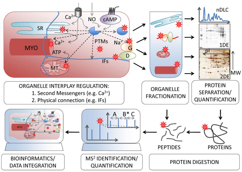
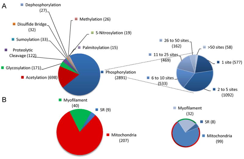
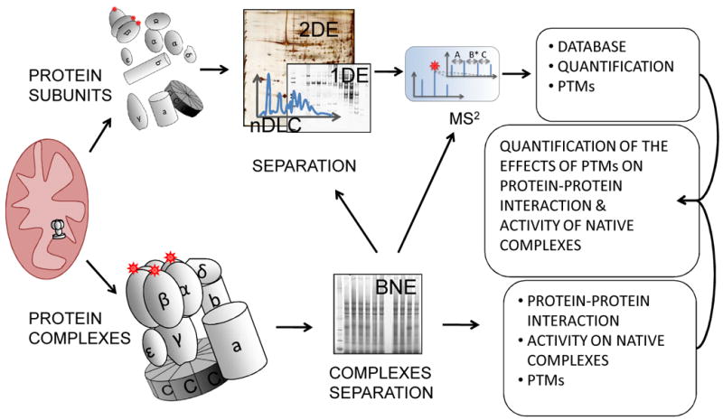
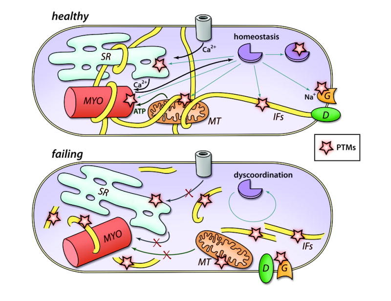
Chronic heart failure is a worldwide cause of mortality and morbidity and is the final outcome of a number of different etiologies. This reflects both the complexity of the disease and our incomplete understanding of its underlying molecular mechanisms. One experimental approach to address this is to study subcellular organelles and how their functions are activated and synchronized under physiological and pathological conditions. In this review, we discuss the application of proteomic technologies to organelles and how this has deepened our perception of the cellular proteome and its alterations with heart failure. The use of proteomics to monitor protein quantity and posttranslational modifications has revealed a highly intricate and sophisticated level of protein regulation. Posttranslational modifications have the potential to regulate organelle function and interplay most likely by targeting both structural and signaling proteins throughout the cell, ultimately coordinating their responses. The potentials and limitations of existing proteomic technologies are also discussed emphasizing that the development of novel methods will enhance our ability to further investigate organelles and decode intracellular communication.


Figures

Figure 1
Figure 1. A proteomic strategy to study organellar communication
After organelle fractionation, proteomic methods including protein separation, quantification and identification are employed to study the different sub-proteomes. Integrated analysis of the data generated from several organelles can help highlighting common molecular components involved in the coordination of different cell compartments. MYO, myofilament; MT, mitochondrion; G, gap junction; D, desmosome.
Figure 2
Figure 2. PTMs in heart proteins
A) Proteins annotated as “heart” with their site specific PTMs* (left) with emphasis on phosphorylated proteins grouped based on the total number of phosphorylation sites (right). B) Proteins annotated as “heart” with their subcellular localization in either SR , sarcomeres or mitochondria; the total number (left) and the number of the ones that are phosphorylated (right) are provided, as reported in the Uniprot Protein Knowledgebase (UniprotKB). *Only PTMs that appeared in more than 15 entries in the Human protein reference database were considered.
Figure 3
Figure 3. Proteomic to study PTM regulation of protein-protein interaction
Proteins and protein complexes can be alternatively separated from organelles, such as mitochondria. As an example, schematically presented, ATP-synthase (complex V, adapted from Elston T, et al., Nature. 1998, 29;391:510-3)can be denatured into its individual subunits or separated as a whole. The first approach is more suitable to monitor PTMs, the latter provides information on complex composition. The integrated analysis of the two strategies can indicate the role of PTMs in complex formation.
Figure 4
Figure 4. Organelle Dyscoordination in Heart Failure
Schematic presentation of features in a failing cardiomyocyte: increased neurohormonal stimulation and mechanical stress affects cellular contractility, energy metabolism, conduction, intracellular signaling and cell structure mislocalization. We propose that dys-regulation of various organelles could be considered another feature of the failing heart. We further hypothesize that this is regulated through changes in the homeostasis of secondmessengers, resulting in dynamic regulation of PTMs, and by the disruption of cytoskeletal structures such as IFs. MYO, myofilament; MT, mitochondrion; G, gap junction; D, desmosome.

References

    1. Diwan A, Dorn GW., 2nd Decompensation of cardiac hypertrophy: Cellular mechanisms and novel therapeutic targets. Physiology (Bethesda) 2007;22:56–64. - PubMed
    1. Houser SR, Margulies KB. Is de pressed myocyte contractility centrally involved in heart failure? Circ Res. 2003;92:350–358. - PubMed
    1. Lehnart SE, Maier LS, Hasenfuss G. Abnormalities of calcium metabolism and myocardial contractility depression in the failing heart. Heart Fail Rev. 2009;14:213–224. - PMC - PubMed
    1. Rosca MG, Hoppel CL. Mitochondria in heart failure. Cardiovasc Res. 2010 - PMC - PubMed
    1. Capetanaki Y. Desmin cytoskeleton: A potential regulator of muscle mitochondrial behavior and function. Trends Cardiovasc Med. 2002;12:339–348. - PubMed

Publication types

MeSH terms