. 2011 May;300(5):H1753-61.
doi: 10.1152/ajpheart.00946.2010. Epub 2011 Feb 18.

S1P lyase: a novel therapeutic target for ischemia-reperfusion injury of the heart

Affiliations

S1P lyase: a novel therapeutic target for ischemia-reperfusion injury of the heart

Padmavathi Bandhuvula et al. Am J Physiol Heart Circ Physiol. 2011 May.

Abstract

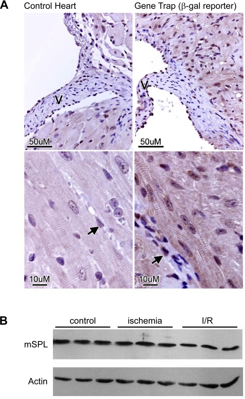
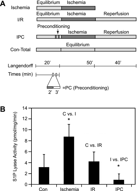
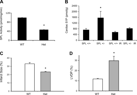
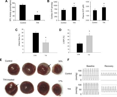
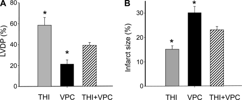
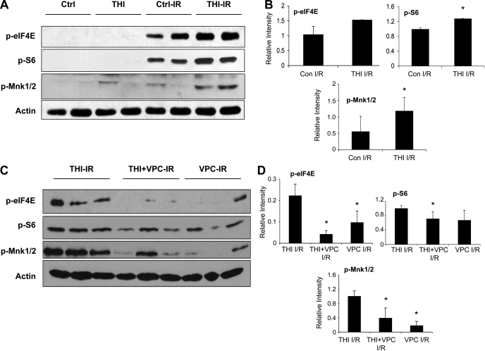
Sphingosine-1-phosphate (S1P) is a bioactive sphingolipid that promotes cardiomyocyte survival and contributes to ischemic preconditioning. S1P lyase (SPL) is a stress-activated enzyme responsible for irreversible S1P catabolism. We hypothesized that SPL contributes to oxidative stress by depleting S1P pools available for cardioprotective signaling. Accordingly, we evaluated SPL inhibition as a strategy for reducing cardiac ischemia-reperfusion (I/R) injury. We measured SPL expression and enzyme activity in murine hearts. Basal SPL activity was low in wild-type cardiac tissue but was activated in response to 50 min of ischemia (n = 5, P < 0.01). Hearts of heterozygous SPL knockout mice exhibited reduced SPL activity, elevated S1P levels, smaller infarct size, and increased functional recovery after I/R compared with littermate controls (n = 5, P < 0.01). The small molecule tetrahydroxybutylimidazole (THI) is a Federal Drug Administration-approved food additive that inhibits SPL. When given overnight at 25 mg/l in drinking water, THI raised S1P levels and reduced SPL activity (n = 5, P < 0.01). THI reduced infarct size and enhanced hemodynamic recovery in response to 50 min of ischemia and to 40 min of reperfusion in ex vivo hearts (n = 7, P < .01). These data correlated with an increase in MAP kinase-interacting serine/threonine kinase 1, eukaryotic translation initiation factor 4E, and ribosomal protein S6 phosphorylation levels after I/R, suggesting that SPL inhibition enhances protein translation. Pretreatment with an S1P₁ and S1P₃ receptor antagonist partially reversed the effects of THI. These results reveal, for the first time, that SPL is an ischemia-induced enzyme that can be targeted as a novel strategy for preventing cardiac I/R injury.


Figures

Fig. 1.
Fig. 1.
Sphingosine-1-phosphate (S1P) lyase (SPL) expression and activity in the murine heart. A: immunohistochemical detection of a β-galactosidase (β-gal) reporter in hearts of SPL gene-trap reporter mice and reporterless littermate controls. β-Gal was detected in the endothelium (arrows), endocardium lining the valve (V), and cardiomyocytes. B: SPL protein expression in wild-type (WT) heart tissue. See text for details. mSPL, murine SPL.
Fig. 2.
Fig. 2.
SPL activity and expression in response to ischemia/reperfusion (I/R) injury in WT hearts. A: I/R protocols. IPC, ischemic preconditioning; Con-Total, 110-min equilibrium normoxic control. B: SPL enzyme activity. n = 5 hearts per treatment. Con and C, control; I, ischemia. *P < 0.05.
Fig. 3.
Fig. 3.
Mouse models with reduced SPL expression are less susceptible to I/R injury. A and B: tissue (intestine) SPL activity (A) and cardiac S1P levels (B) in mice heterozygous (Het) for a gene-trap targeted disruption of Sgpl1 and littermate controls. C and D: infarct size (C) and left ventricular (LV) developed pressure (LVDP; D) after I/R. Four WT and 5 heterozygous mouse hearts were used. *P < 0.05.
Fig. 4.
Fig. 4.
Tetrahydroxybutylimidazole (THI) pretreatment protects against I/R injury of hearts from WT mice. A and B: tissue (thymus) SPL activity (A) and cardiac and plasma S1P levels (B) in vehicle- and THI-treated mice. C: infarction size in control and THI-treated mice as percentage of the area at risk, which is the entire LV in this global ischemia model. Seven control mice and six THI-treated mice were used. D: LVDP at the end of reperfusion. E: typical infarcts in control and THI-treated hearts. Arrows point to representative infarct areas. F: representative examples of LVDP in control and THI-treated hearts. As shown, the developed pressure was substantially greater in the THI-treated heart at the end of reperfusion. See D for group details. *Statistical significance (P < 0.05).
Fig. 5.
Fig. 5.
The S1P cell surface receptor antagonist VPC-23019 (VPC) reduces the effect of THI. Mouse hearts were subjected to I/R injury as described in methods (n = 4 hearts/group). A: compared with THI, the S1P antagonist VPC had no significant effect on LVDP at the end of reperfusion. Values, expressed as a percentage of baseline, were similar to those obtained in the absence of VPC (compare with Fig. 4D). In hearts harvested from THI-treated mice, the results were intermediate. B: Infarct size, expressed as a percentage of the risk area, which is the entire LV in this global ischemia model, was reduced by treatment with THI, and VPC alone had no effect on the extent of infarction (compare with Fig. 4C). However, in the presence of VPC, the results in THI-treated mice were intermediate. These data establish S1P receptor involvement but also suggest a possible intracellular role for S1P. Statistics were determined by one-way ANOVA with post hoc Student-Newman-Keuls testing. *P < 0.05 vs. all other conditions.
Fig. 6.
Fig. 6.
THI treatment potentiates the activation of protein translation after I/R in WT mouse hearts. A: phosphorylated eukaryotic translation initiation factor 4E (p-eIF4E), phosphorylated ribosomal S6 protein (p-S6), phosphorylated MAPK-interacting serine/threonine kinase 1/2 (p-Mnk), and actin levels in heart tissues from THI-treated and control (Ctrl) mice before and after I/R. B: ImageJ quantification of post-I/R signals normalized to actin. These blots are representative of 4 separate experiments. *P < 0.05 vs. control I/R. C: p-eIF4E, p-S6, p-Mnk, and actin levels in tissues from THI-, VPC-, and THI + VPC-treated hearts after I/R. D: ImageJ quantification of the results shown in C. *P < 0.05 vs. THI I/R.

References

    1. Bagdanoff J, Donoviel M, Nouraldeen A, Tarver J, Fu Q, Carlsen M, Jessop T, Zhang H, Hazelwood J, Nguyen H, Baugh S, Gardyan M, Terranova K, Barbosa J, Yan J, Bednarz M, Layek S, Courtney L, Taylor J, Digeorge-Foushee A, Gopinathan S, Bruce D, Smith T, Moran L, O'Neill E, Kramer J, Lai Z, Kimball S, Liu Q, Sun W, Yu S, Swaffield J, Wilson A, Main A, Carson K, Oravecz T, Augeri D. Inhibition of sphingosine-1-phosphate lyase for the treatment of autoimmune disorders. J Med Chem 52: 3941– 3953, 2009 - PubMed
    1. Bandhuvula P, Fyrst H, Saba J. A rapid fluorescent assay for sphingosine-1-phosphate lyase enzyme activity. J Lipid Res 48: 2769– 2778, 2007 - PubMed
    1. Bektas M, Allende ML, Lee BG, Chen W, Amar MJ, Remaley AT, Saba JD, Proia RL. Sphingosine 1-phosphate lyase deficiency disrupts lipid homeostasis in liver. J Biol Chem 285: 10880– 10889, 2010 - PMC - PubMed
    1. Fyrst H, Saba J. An update on sphingosine-1-phosphate and other sphingolipid mediators. Nat Chem Biol 6: 489– 497, 2010 - PMC - PubMed
    1. Hait NC, Allegood J, Maceyka M, Strub GM, Harikumar KB, Singh SK, Luo C, Marmorstein R, Kordula T, Milstien S, Spiegel S. Regulation of histone acetylation in the nucleus by sphingosine-1-phosphate. Science 325: 1254– 1257, 2009 - PMC - PubMed

Publication types

LinkOut - more resources