. 2011 Apr 1;186(7):4443-54.
doi: 10.4049/jimmunol.1002449. Epub 2011 Feb 18.

β2-adrenergic receptor activation prevents rodent dopaminergic neurotoxicity by inhibiting microglia via a novel signaling pathway

Affiliations

β2-adrenergic receptor activation prevents rodent dopaminergic neurotoxicity by inhibiting microglia via a novel signaling pathway

Li Qian et al. J Immunol. .

Abstract

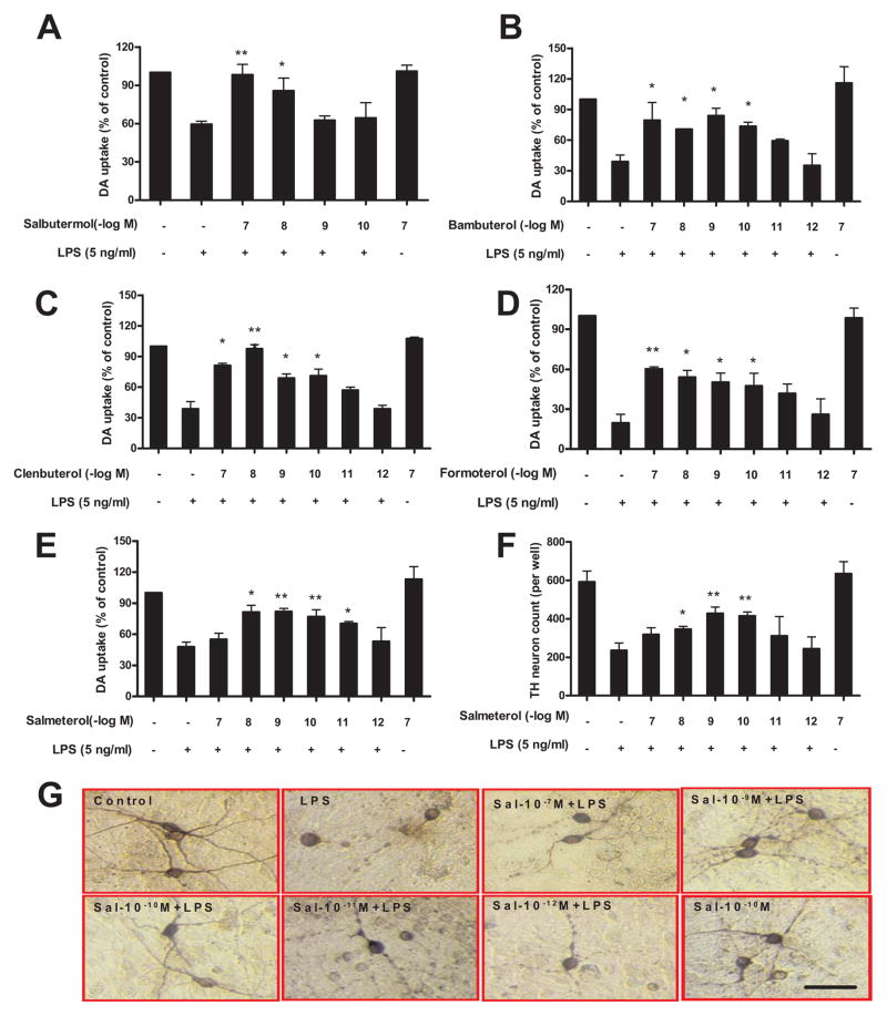
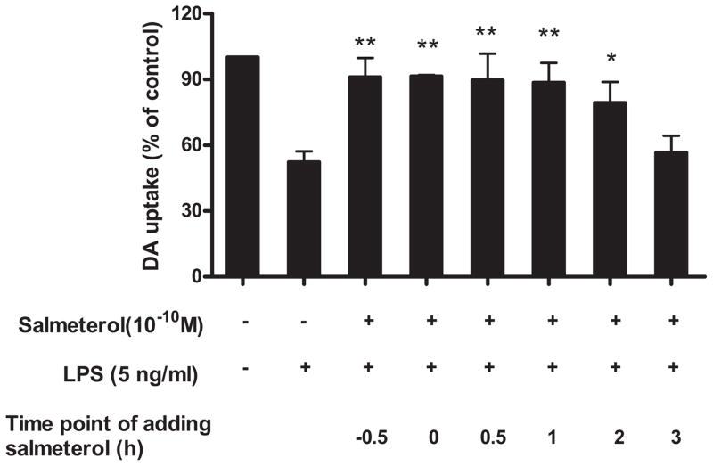
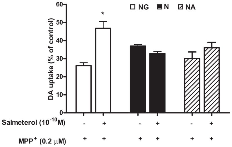
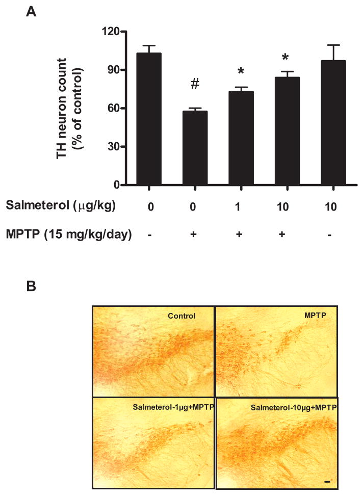
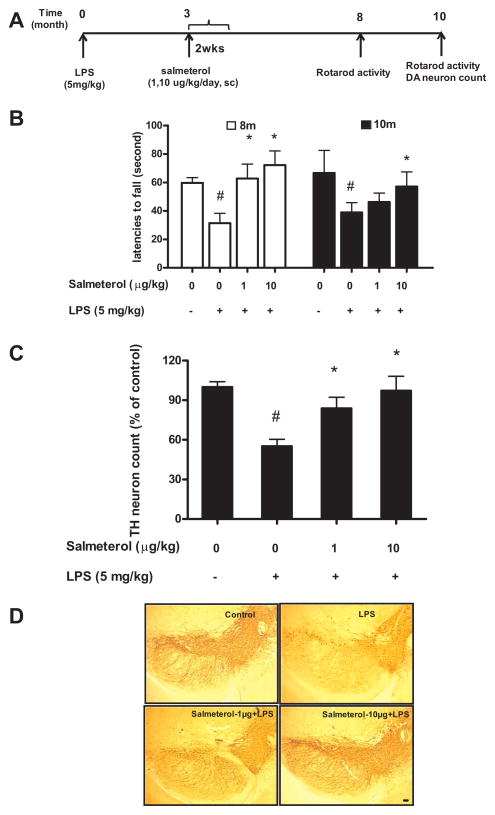
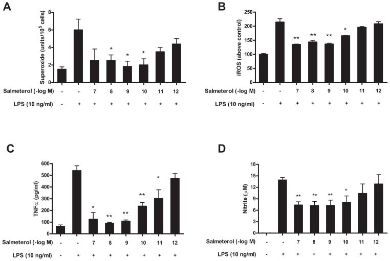
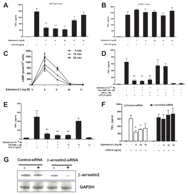
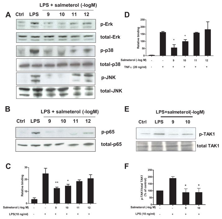
The role of the β2 adrenergic receptor (β2AR) in the regulation of chronic neurodegenerative inflammation within the CNS is poorly understood. The purpose of this study was to determine neuroprotective effects of long-acting β2AR agonists such as salmeterol in rodent models of Parkinson's disease. Results showed salmeterol exerted potent neuroprotection against both LPS and 1-methyl-4-phenyl-1,2,3,6-tetrahydropyridine/1-methyl-4-phenylpyridinium-induced dopaminergic neurotoxicity both in primary neuron-glia cultures (at subnanomolar concentrations) and in mice (1-10 μg/kg/day doses). Further studies demonstrated that salmeterol-mediated neuroprotection is not a direct effect on neurons; instead, it is mediated through the inhibition of LPS-induced microglial activation. Salmeterol significantly inhibited LPS-induced production of microglial proinflammatory neurotoxic mediators, such as TNF-α, superoxide, and NO, as well as the inhibition of TAK1-mediated phosphorylation of MAPK and p65 NF-κB. The anti-inflammatory effects of salmeterol required β2AR expression in microglia but were not mediated through the conventional G protein-coupled receptor/cAMP pathway. Rather, salmeterol failed to induce microglial cAMP production, could not be reversed by either protein kinase A inhibitors or an exchange protein directly activated by cAMP agonist, and was dependent on β-arrestin2 expression. Taken together, our results demonstrate that administration of extremely low doses of salmeterol exhibit potent neuroprotective effects by inhibiting microglial cell activation through a β2AR/β-arrestin2-dependent but cAMP/protein kinase A-independent pathway.


Conflict of interest statement

Disclosures

The authors have no financial conflict of interest.

Figures

FIGURE 1
FIGURE 1
Low doses of β2AR agonists protect DA neurons against LPS-induced toxicity. Rat primary mesencephalic neuron-glia cultures were seeded in a 24-well culture plate at 5×10⁵, then pretreated with vehicle or indicated concentrations of β2AR agonists, including short-acting β2AR agonist salbutermol (A), long-acting β2AR agonists bambuterol (B), clenbuterol (C), formoterol (D) and salmeterol (E–G) for 30 min before the addition of 5 ng/ml LPS. Seven days later, the LPS-induced DA neurotoxicity was quantified by the [3H]-DA uptake assay (A–E); the immunocytochemical analysis of salmeterol and LPS treated cells, including TH-IR neuron counts (F) and the representative pictures of TH+ neuron immunostaining (G). Results in A–E were expressed as a percentage of the vehicle-treated control cultures and were the means ± SE. from three independent experiments in triplicate. *P<0.05, **P<0.01 compared with the LPS-treated cultures. Scale bar: 50 μm.
FIGURE 2
FIGURE 2
Salmeterol mediates neuroprotection when administered prior to or after exposure to LPS. Rat primary mesencephalic neuron-glia cultures were seeded in a 24-well culture plate at 5×10⁵, vehicle or salmeterol at 10−10 M was added to neuron-glia cultures 30 min prior to, simultaneously with, 30 min, 1, 2, or 3 hrs following the addition of LPS. DA neurotoxicity was measured 7 days later. Results were expressed as a percentage of the vehicle-treated control cultures and were the means ± SE from 3 independent experiments in triplicate. *P<0.05, **P<0.01 compared with the LPS-treated cultures.
FIGURE 3
FIGURE 3
Salmeterol mediated neuroprotection is microglia dependent. Mesencephalic midbrain neuron-glia cultures (open bars, NG, which contain ~10% microglia), neuron-enriched cultures (solid bars, N, which contain <0.1% microglia) and neuron-astrocyte cultures (cross-hatched bars, NA, which contain <0.1% microglia) were pretreated with either vehicle or 10−10 M salmeterol for 30 min prior to the addition of 0.2 μM MPP+. The [3H]-DA uptake measurements were performed 7 days following MPP+ treatment. Results were expressed as a percentage of the vehicle-treated control cultures and were the means ± SE from 3 independent experiments in triplicate. * P<0.05 compared with the MPP+-treated cultures.
FIGURE 4
FIGURE 4
Salmeterol was also capable of inhibiting progressive DA neurodegeneration induced by injection of the neurotoxin MPTP. To induce DA neurodegeneration, MPTP (15 mg/kg) was injected s.c. into C57BL/6J mice for 6 consecutive days. Experimental mice (8 mice/group) were also given either saline or doses of salmeterol at 1μg/kg/day or at 10μg/kg/day by continuous infusion (via an Alzet mini-pump) for 2 weeks starting two days before the first MPTP injection. We then assessed the survival of DA neurons in the SNpc 21 days following the last MPTP injection through quantification of TH-positive neurons (A). Representative pictures of TH-IR neurons staining for different treatment groups are shown (B). *P<0.05, compared with the LPS-treated mice. #P<0.05, compared with saline-treated mice. Scale bar: 50 μm.
FIGURE 5
FIGURE 5
Salmeterol significantly attenuates the loss of rotarod activity and DA neurons in SNpc induced by systemic LPS injection. C57BL/6J mice received single LPS injection (5mg/kg, i.p), and 3 months later, mice were infused with either vehicle alone or salmeterol (1 or 10 μg/kg/day) for 2 weeks (via an Alzet mini-pump). Eight months and 10 months after LPS injection, rotarod tests were performed (A–B), and then at 10 months mice were sacrificed, brains were harvested and sectioned, and 24 sections (rostral to caudal: 4.52–5.36 mm posterior to bregma) were collected as described in methods. Eight evenly spaced brain sections from vehicle, LPS-injected, or LPS-injected salmeterol-treated animals were immunostained with anti-TH antibody, and the number of TH-IR neurons in the SN was counted. Data were expressed as percent loss compared to saline-injected controls (C). Representative pictures of TH-IR neurons staining for different treatment groups are shown (D). *P<0.05,compared with the LPS-treated mice. #P<0.05, compared with saline-treated mice. Scale bar: 50 μm.
FIGURE 6
FIGURE 6
Effect of salmeterol on LPS-induced production of pro-inflammatory factors from microglia. Microglia-enriched cultures were seeded at a density of 1×10⁵/well. Cells were pretreated with vehicle or various concentrations of salmeterol for 30 min followed by the addition of LPS. The production of LPS-induced extracellular superoxide production (A) was measured as SOD-inhabitable reduction of WST-1. LPS-induced intracellular ROS was determined by probe DCFH-DA (B). Salmererol’s effect on LPS-induced production of TNFα and nitrite were shown in Fig. 6C and Fig. 6D. Results were expressed as mean ± SE from three to five independent experiments in triplicate. *P<0.05, **P<0.01 compared with the LPS-treated cultures.
FIGURE 7
FIGURE 7
Low dose salmeterol-mediated anti-inflammatory effects are β2AR/β-arrestin2 dependent, but cAMP/PKA and cAMP/EPAC-independent. Microglia culture prepared from C57/BL6 (A) or β2AR deficient mice (B) were pretreated with vehicle or indicated concentrations of salmeterol for 30 min prior to the addition of LPS. Supernatant were collected at 3h after LPS addition for TNFα analysis (A–B). Figure C: Enriched primary microglia were incubated with vehicle or indicated concentrations of salmeterol at 37°C for 5, 10, or 30 min. After incubation, the cells were lysed and cAMP levels were determined using a cAMP assay kit. Data were expressed as pmol cAMP per 1 million cells (C). Figures D and E: Enriched primary microglia cells were pretreated with vehicle or PKA inhibitors, including H89 (1 μM for 45 min), Rp-cAMP (50 μM for 45 min) (D), or EPAC agonist 8CPT-2′-O-Me-cAMP (CPTOMe) (10 μM for 45 min) (E) prior to stimulation with salmeterol (10−10M) and LPS (5 ng/ml). Supernatants were collected 3h after LPS addition to measure TNFα levels. Figure F and G: Primary microglia cells were transfected with 100 pmol specific β-arrestin2 siRNA or control siRNA, and 48 hrs after transfection cells were treated with indicated concentrations of salmeterol for 30 min prior to addition of LPS. Supernatants were collected for TNFα assay (F). Knockdown of expression of the β-arrestin2 was determined by western blot analysis (G). Results in A–F were expressed as mean ± SE from three to four independent experiments in triplicate. *P<0.05, **P<0.01 compared with the LPS-treated cultures.
FIGURE 8
FIGURE 8
Salmeterol significantly suppresses LPS-induced MAPK and NF-κB activation through inhibition of TAK-1 phosphorylation. Enriched microglia were pretreated with vehicle or salmeterol (10−9–10−12 M) for 30 min followed by treatment with LPS (10 ng/ml) for 15 min. Cells were then harvested, and the amounts of phosphorylated and total MAPK (ERK1/2, p38, and JNK1/2) (A) and phosphorylated and total NF-κB p65 (B) were determined by western blot analysis, respectively. Representative western blots for ERK1/2, p38, JNK and p65 phosphorylation are shown from 3 independent experiments. Figures C and D: primary microglia were pretreated with salmeterol (10−9–10−12 M) for 30 min, then treated with LPS (C) or TNFα (D) for 30 min. Nuclear extracts were prepared from these cells, and NF-κB (p65) DNA binding activity was detected using the NF-κB (p65) Transcription Factor Assay Kit. Figure E and F: Enriched microglia were pretreated with vehicle or salmeterol (10−9–10−10 M) for 30 min followed by the treatment with LPS (10 ng/ml) for 15 min, cells were then harvested, and the amounts of phosphorylated TAK1 and total TAK1 are show in western blot analysis using specific antibodies (E). ImageJ software was used to quantitate the intensity of the phosphorylated TAK1 and total TAK1 bands in western blot, and the results given in figure F represents the percentage difference of the ratio of phosphorylated TAK1 compared with total TAK1 normalized to the vehicle-treated control (F).

References

    1. Koff WC, Fann AV, Dunegan MA, Lachman LB. Catecholamine-induced suppression of interleukin-1 production. Lymphokine Res. 1986;5:239–247. - PubMed
    1. van der Poll T, Jansen J, Endert E, Sauerwein HP, van Deventer SJ. Noradrenaline inhibits lipopolysaccharide-induced tumor necrosis factor and interleukin 6 production in human whole blood. Infect Immun. 1994;62:2046–2050. - PMC - PubMed
    1. Sekut L, Champion BR, Page K, Menius JA, Jr, Connolly KM. Anti-inflammatory activity of salmeterol: down-regulation of cytokine production. Clin Exp Immunol. 1995;99:461–466. - PMC - PubMed
    1. Severn A, Rapson NT, Hunter CA, Liew FY. Regulation of tumor necrosis factor production by adrenaline and beta-adrenergic agonists. J Immunol. 1992;148:3441–3445. - PubMed
    1. Farmer P, Pugin J. beta-adrenergic agonists exert their “anti-inflammatory” effects in monocytic cells through the IkappaB/NF-kappaB pathway. Am J Physiol Lung Cell Mol Physiol. 2000;279:L675–682. - PubMed

Publication types

MeSH terms