. 2011 Apr 15;286(15):12912-23.
doi: 10.1074/jbc.M110.143693. Epub 2011 Feb 18.

Collagen I induces discoidin domain receptor (DDR) 1 expression through DDR2 and a JAK2-ERK1/2-mediated mechanism in primary human lung fibroblasts

Affiliations

Collagen I induces discoidin domain receptor (DDR) 1 expression through DDR2 and a JAK2-ERK1/2-mediated mechanism in primary human lung fibroblasts

Pedro A Ruiz et al. J Biol Chem. .

Abstract

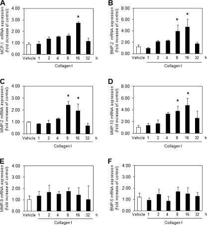
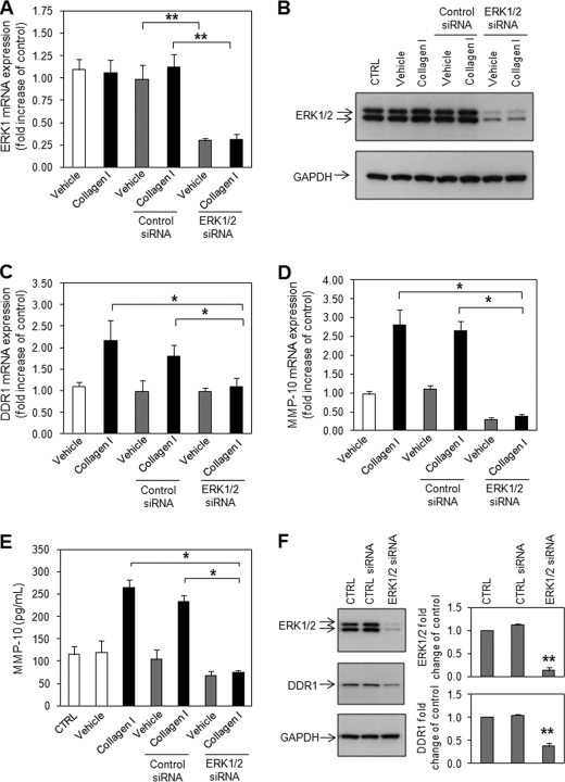
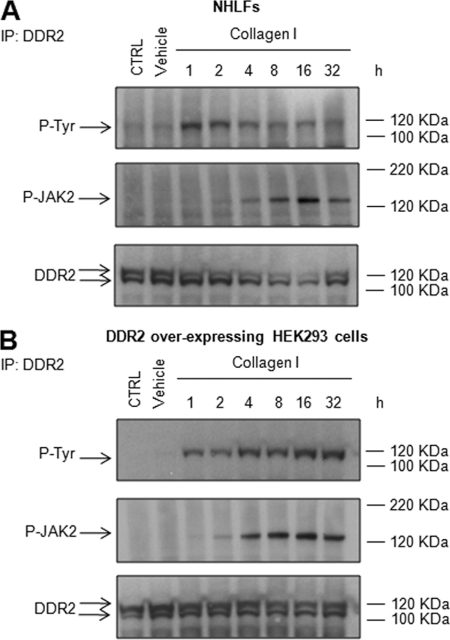
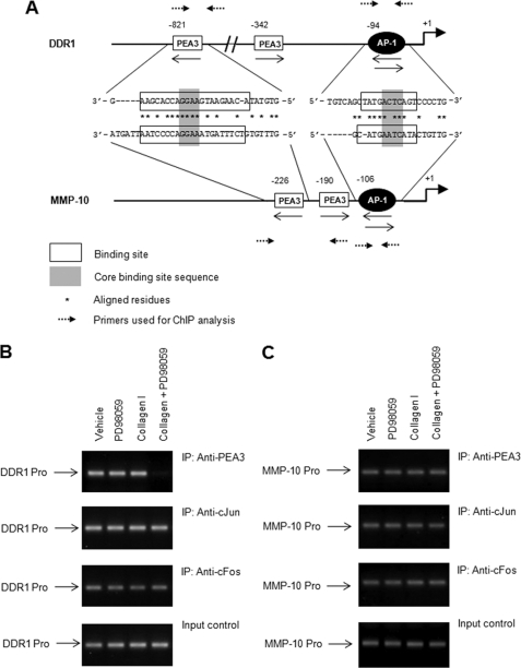
Discoidin domain receptors (DDRs) DDR1 and DDR2 are receptor tyrosine kinases with the unique ability among receptor tyrosine kinases to respond to collagen. Several signaling molecules have been implicated in DDR signaling, including Shp-2, Src, and MAPK pathways, but a detailed understanding of these pathways and their transcriptional targets is still lacking. Similarly, the regulation of the expression of DDRs is poorly characterized with only a few inflammatory mediators, such as lipopolysaccharide and interleukin-1β identified as playing a role in DDR1 expression. DDRs have been reported to induce the expression of various genes including matrix metalloproteinases and bone morphogenetic proteins, but the regulatory mechanisms underlying DDR-induced gene expression remain to be determined. The aim of the present work was to elucidate the molecular mechanisms implicated in the expression of DDRs and to identify DDR-induced signaling pathways and target genes. Our data show that collagen I induces the expression of DDR1 in a dose- and time-dependent manner in primary human lung fibroblasts. Furthermore, activation of DDR2, JAK2, and ERK1/2 MAPK signaling pathways was essential for collagen I-induced DDR1 and matrix metalloproteinase 10 expression. Finally, inhibition of the ERK1/2 pathway abrogated DDR1 expression by blocking the recruitment of the transcription factor polyoma enhancer A-binding protein 3 to the DDR1 promoter. Our data provide new insights into the molecular mechanisms of collagen I-induced DDR1 expression and demonstrate an important role for ERK1/2 activation and the recruitment of polyoma enhancer-A binding protein 3 to the DDR1 promoter.


Figures

FIGURE 1.
FIGURE 1.
Collagen I but not collagen IV induces DDR1 expression in a dose-dependent manner in NHLFs. A and B, NHLFs were serum-starved overnight and incubated with vehicle (AcH, 0.1 m) or the specified concentrations of collagen I and collagen IV for 16 h. Total RNA was isolated and reverse transcribed, and real time quantitative PCR was performed using the TaqMan system with specific primers and TaqMan Probes for human DDR1 (A), DDR2 (B), and GAPDH. The expression changes (fold increases) were calculated relative to unstimulated control cells after normalizing with GAPDH. The results are representative of mean fold increases ± S.D. of three independent experiments done in triplicate (n = 9; *, p < 0.05). C, NHLFs were serum-starved overnight and incubated with vehicle (AcH, 0.1 m) or collagen I (25 μg/ml) for 16 h. The cells were collected using 5 mm EDTA and incubated with biotinylated goat anti-DDR1 antibody or isotype control biotinylated normal goat IgG, followed by phycoerythrin (PE)-conjugated streptavidin. Flow cytometry was performed using FACSCanto. The frequency of positive results in the selected population is shown. The results are representative of mean fold increases ± S.D. of two independent experiments done in triplicate (n = 6). D, total RNA was isolated from NHLFs and reverse transcribed, and real time quantitative PCR was performed using the TaqMan system with specific primers for human DDR1, DDR1a, DDR1b, DDR1c, DDR1d, DDR1e, and DDR2. PCR products were run on 2% agarose gel. E, NHLFs were serum-starved overnight and incubated with collagen I (25 μg/ml) or vehicle (AcH, 0.1 m) at various time points. Real time quantitative PCR was performed as described above. The results are representative of mean fold increases ± S.D. of three independent experiments done in triplicate (n = 9; *, p < 0.05).
FIGURE 2.
FIGURE 2.
Collagen I induces MCP-1, BMP-2, MMP-2, and MMP-10 mRNA expression in a time-dependent manner in NHLFs. NHLFs were serum-starved overnight and incubated with collagen I (25 μg/ml) or vehicle (AcH, 0.1 m) at various time points. Total RNA was isolated and reverse transcribed, and real time quantitative PCR was performed using the TaqMan system with specific primers and TaqMan Probes for human MCP-1 (A), BMP-2 (B), MMP-2 (C), MMP-10 (D), MMP-9 (E), BMP-5 (F), and GAPDH. The expression changes (fold increases) were calculated relative to unstimulated control cells after normalizing with GAPDH. The results are representative of mean fold increases ± S.D. of three independent experiments done in triplicate (n = 9; *, p < 0.05).
FIGURE 3.
FIGURE 3.
Collagen I induces DDR1 and MMP-10 expression through DDR and independently of β1 integrin in NHLFs. A, B, and D, NHLFs were serum-starved overnight. The cells were then incubated with AMN107 (1 mm) or anti-human β1 integrin antibody (10 μg/ml) for 1 h prior to 16 h stimulation with collagen I (25 μg/ml). Total RNA was isolated and reverse transcribed, and real time quantitative PCR was performed using the TaqMan system with specific primers and TaqMan Probes for human DDR1 (A), MMP-10 (B), Cyclin D1 (D), and GAPDH. The expression changes (fold increases) were calculated relative to unstimulated control (CTRL) cells after normalizing with GAPDH. The results are representative of mean fold increases ± S.D. of three independent experiments done in triplicate (n = 9; *, p < 0.05; **, p < 0.01). C, MMP-10 protein was measured in the culture supernatant of stimulated NHLFs by ELISA. The results are representative of mean fold increases ± S.D. of three independent experiments done in triplicate (n = 9; *, p < 0.05).
FIGURE 4.
FIGURE 4.
Collagen I induces DDR1 and MMP-10 mRNA expression in HEK293 cells overexpressing DDR2 but not in DDR1a or DDR1b. HEK293 cells overexpressing DDR1a, DDR1b, or DDR2 were serum-starved overnight, and the cells were then incubated for 16 h with collagen I (25 μg/ml). Total RNA was isolated and reverse transcribed, and real time quantitative PCR was performed using the TaqMan system with specific primers and TaqMan Probes for human DDR1 (A), MMP-10 (B), and GAPDH. The expression changes (fold increases) were calculated relative to unstimulated control cells after normalizing with GAPDH. The results are representative of mean fold increases ± S.D. of two independent experiments done in triplicate (n = 6; *, p < 0.05; **, p < 0.01).
FIGURE 5.
FIGURE 5.
Collagen I induces MMP-10 mRNA expression by a JAK2-ERK1/2-dependent mechanism in NHLFs and DDR2-overexpressing HEK293 cells. A and B, NHLFs (A) and DDR2-overexpressing HEK293 cells (B) were serum-starved overnight. The cells were then incubated with AG490 (25 μm), PD98059 (20 μm), or SB203580 (20 μm) for 1 h prior to 16 h of stimulation with collagen I (25 μg/ml). Total RNA was isolated and reverse transcribed, and real time quantitative PCR was performed using the TaqMan system with specific primers and TaqMan Probes for human MMP-10 and GAPDH. The expression changes (fold increases) were calculated relative to unstimulated control (CTRL) cells after normalizing with GAPDH. The results are representative of mean fold increases ± S.D. of three independent experiments done in triplicate (n = 9; *, p < 0.05). C, DDR2-overexpressing HEK293 cells were serum-starved overnight and incubated with collagen I (25 μg/ml) at various time points. Total protein was isolated and subjected to SDS-PAGE followed by anti-phospho-JAK2, anti-JAK2, anti-phospho-ERK1/2, anti-ERK1/2 MAPK, and anti-DDR1 immunoblotting. Anti-GAPDH was used as loading control. Quantification relative to control cells after normalization to GAPDH or total protein is presented. The results are representative of mean fold increase ± S.D. of three independent experiments (n = 3; *, p < 0.05; **, p < 0.01). D and E, DDR2-overexpressing HEK293 cells were serum-starved overnight. The cells were then incubated with AG490 (25 μm) (D) or PD98059 (20 μm) (E) for 1 h prior to 16 h of stimulation with collagen I (25 μg/ml). Total protein was isolated and subjected to SDS-PAGE followed by anti-phospho-JAK2, anti-JAK2, anti-phospho-ERK1/2 MAPK, anti-ERK1/2 MAPK, and anti-DDR1 immunoblotting. Anti-GAPDH was used as loading control. Quantification relative to control cells after normalization to GAPDH or total protein is presented. The results are representative of mean fold increase ± S.D. of three independent experiments (n = 3; *, p < 0.05; **, p < 0.01; ***, p < 0.001).
FIGURE 6.
FIGURE 6.
Collagen I induces DDR1 and MMP-10 expression through DDR2 in NHLFs. A, C, and D, NHLFs were reverse transfected with DDR2-specific siRNA or negative control (CTRL) siRNA using Lipofectamine RNAiMAX. 48 h after transfection, NHLFs were serum-starved for 24 h and incubated with collagen I (25 μg/ml) or vehicle (AcH, 0.1 m) for 16 h. Total RNA was isolated and reverse transcribed, and real time quantitative PCR was performed using the TaqMan system with specific primers and TaqMan Probes for human DDR2 (A), DDR1 (C), MMP-10 (D), and GAPDH. The expression changes (fold increases) were calculated relative to unstimulated control cells after normalizing with GAPDH. The results are representative of mean fold increases ± S.D. of two independent experiments done in triplicate (n = 6; *, p < 0.05; **, p < 0.01). B, NHLFs were reverse transfected with 10 nm DDR2-specific siRNA or negative control siRNA. 48 h after transfection, NHLFs were serum-starved for 24 h and incubated with collagen I (25 μg/ml) or vehicle (AcH, 0.1 m) for 16 h. Total protein was isolated and subjected to SDS-PAGE followed by anti-DDR2 immunoblotting. Anti-GAPDH was used as loading control. The results are representative of three independent experiments. E, NHLFs were transfected with DDR2-specific siRNA or negative control siRNA prior to starvation and 16 h of collagen I stimulation. MMP-10 protein was measured in the culture supernatant by ELISA. The results are representative of mean fold increases ± S.D. of two independent experiments done in triplicate (n = 6; *, p < 0.05). F, NHLFs were reverse transfected with DDR2-specific siRNA or negative control siRNA. Total protein was isolated and subjected to SDS-PAGE followed by anti-DDR2, anti-DDR1, anti-phospho-JAK2, anti-JAK2, anti-phospho-ERK1/2, and anti-ERK1/2 immunoblotting. Anti-GAPDH was used as loading control. Quantification relative to control cells after normalization to GAPDH or total protein is presented. The results are representative of mean fold increase ± S.D. of three independent experiments (n = 3; *, p < 0.05; **, p < 0.01).
FIGURE 7.
FIGURE 7.
Collagen I-induced DDR1 and MMP-10 expression is mediated by JAK2 in NHLFs. A, C, and D, NHLFs were reverse transfected with JAK2-specific siRNA or negative control (CTRL) siRNA using Lipofectamine RNAiMAX. 48 h after transfection, NHLFs were serum-starved for 24 h and incubated with collagen I (25 μg/ml) or vehicle (AcH, 0.1 m) for 16 h. Total RNA was isolated and reverse transcribed, and real time quantitative PCR was performed using the TaqMan system with specific primers and TaqMan probes for human JAK2 (A), DDR1 (C), MMP-10 (D), and GAPDH. The expression changes (fold increases) were calculated relative to unstimulated control cells after normalizing with GAPDH. The results are representative of mean fold increases ± S.D. of two independent experiments done in triplicate (n = 6; *, p < 0.05). B, NHLFs were reverse transfected with 10 nm JAK2-specific siRNA or negative control siRNA. 48 h after transfection, NHLFs were serum-starved for 24 h and incubated with collagen I (25 μg/ml) or vehicle (AcH, 0.1 m) for 16 h. Total protein was isolated and subjected to SDS-PAGE followed by anti-JAK2 immunoblotting. Anti-GAPDH was used as loading control. The results are representative of three independent experiments. E, NHLFs were transfected with JAK2-specific siRNA or negative control siRNA prior to starvation and 16 h of collagen I stimulation. MMP-10 protein was measured in the culture supernatant by ELISA. The results are representative of mean fold increases ± S.D. of two independent experiments done in triplicate (n = 6; *, p < 0.05). F, NHLFs were reverse transfected with JAK2-specific siRNA or negative control siRNA. Total protein was isolated and subjected to SDS-PAGE followed by anti-JAK2 and anti-DDR1 immunoblotting. Anti-GAPDH was used as loading control. Quantification relative to control cells after normalization to GAPDH or total protein is presented. The results are representative of mean fold increases ± S.D. of three independent experiments (n = 3; *, p < 0.05; **, p < 0.01).
FIGURE 8.
FIGURE 8.
Collagen I-induced DDR1 and MMP-10 expression is mediated by ERK1/2 in NHLFs. A, C, and D, NHLFs were reverse transfected with ERK1/2-specific siRNA or negative control siRNA using Lipofectamine RNAiMAX. 48 h after transfection, NHLFs were serum-starved for 24 h and incubated with collagen I (25 μg/ml) or vehicle (AcH, 0.1 m) for 16 h. Total RNA was isolated and reverse transcribed, and real time quantitative PCR was performed using the TaqMan system with specific primers and TaqMan probes for human ERK1 (A), DDR1 (C), MMP-10 (D), and GAPDH. The expression changes (fold increases) were calculated relative to unstimulated control cells after normalizing with GAPDH. The results are representative of mean fold increases ± S.D. of two independent experiments done in triplicate (n = 6; *, p < 0.05; **, p < 0.01). B, NHLFs were reverse transfected with 10 nm ERK1/2-specific siRNA or negative control (CTRL) siRNA. 48 h after transfection, NHLFs were serum-starved for 24 h and incubated with collagen I (25 μg/ml) or vehicle (AcH, 0.1 m) for 16 h. Total protein was isolated and subjected to SDS-PAGE followed by anti-ERK1/2 immunoblotting. Anti-GAPDH was used as loading control. The results are representative of three independent experiments. E, NHLFs were transfected with ERK1/2-specific siRNA or negative control siRNA prior to starvation and 16 h of collagen I stimulation. MMP-10 protein was measured in the culture supernatant by ELISA. The results are representative of mean fold increases ± S.D. of two independent experiments done in triplicate (n = 6; *, p < 0.05). F, NHLFs were reverse transfected with ERK1/2-specific siRNA or negative control siRNA. Total protein was isolated and subjected to SDS-PAGE followed by anti-ERK1/2 and anti-DDR1 immunoblotting. Anti-GAPDH was used as loading control. Quantification relative to control cells after normalization to GAPDH or total protein is presented. The results are representative of mean fold increases ± S.D. of three independent experiments (n = 3; *, p < 0.05; **, p < 0.01).
FIGURE 9.
FIGURE 9.
Collagen I induces DDR2 tyrosine phosphorylation and recruitment of phospho-JAK2 to DDR2 in NHLFs and DDR2-overexpressing HEK293 cells. NHLFs (A) and DDR2-overexpressing HEK293 cells (B) were serum-starved overnight and incubated with collagen I (25 μg/ml) at various time points. Total protein was isolated and incubated overnight with anti-DDR2 antibody followed by incubation with A/G-agarose beads for 1 h. Protein was subjected to SDS-PAGE followed by anti-phosphotyrosine and anti-phospho-JAK2 immunoblotting. Anti-DDR2 was used as loading control (CTRL). The results are representative of two independent experiments. IP, immunoprecipitation.
FIGURE 10.
FIGURE 10.
The recruitment of PEA3 to the promoter-binding site of DDR1 is ERK1/2 MAPK-dependent. A, putative binding sites for AP-1 and PEA3 were found in the DDR1 promoter using Genomatix software tools. DDR2-overexpressing HEK293 cells were serum-starved overnight. The cells were then incubated with PD98059 (20 μm) for 1 h prior to 16 h of stimulation with collagen I (25 μg/ml) or vehicle (AcH, 0.1 m). ChIP analysis was performed using antibodies against PEA3 and the two heterodimers of AP-1, c-Jun and c-Fos, for immunoprecipitation (IP). B and C, real time quantitative PCR was performed using the TaqMan system with specific primers and TaqMan Probes for the DDR1 (B) and MMP-10 (C) promoter-binding sites of the nuclear factors PEA3 and AP-1. Aliquots taken prior to immunoprecipitation were used as input control. PCR products were run on 2% agarose gel. The results are representative of two independent experiments.

References

    1. Alves F., Vogel W., Mossie K., Millauer B., Höfler H., Ullrich A. (1995) Oncogene 10, 609–618 - PubMed
    1. Labrador J. P., Azcoitia V., Tuckermann J., Lin C., Olaso E., Mañes S., Brückner K., Goergen J. L., Lemke G., Yancopoulos G., Angel P., Martínez C., Klein R. (2001) EMBO Rep. 2, 446–452 - PMC - PubMed
    1. Vogel W., Gish G. D., Alves F., Pawson T. (1997) Mol. Cell 1, 13–23 - PubMed
    1. Shrivastava A., Radziejewski C., Campbell E., Kovac L., McGlynn M., Ryan T. E., Davis S., Goldfarb M. P., Glass D. J., Lemke G., Yancopoulos G. D. (1997) Mol. Cell 1, 25–34 - PubMed
    1. Vogel W. F., Abdulhussein R., Ford C. E. (2006) Cell Signal. 18, 1108–1116 - PubMed

Publication types

MeSH terms

LinkOut - more resources