. 2011 Mar;10(3):243-51.
doi: 10.1038/nmat2960. Epub 2011 Feb 20.

Interbilayer-crosslinked multilamellar vesicles as synthetic vaccines for potent humoral and cellular immune responses

Affiliations

Interbilayer-crosslinked multilamellar vesicles as synthetic vaccines for potent humoral and cellular immune responses

James J Moon et al. Nat Mater. 2011 Mar.

Abstract

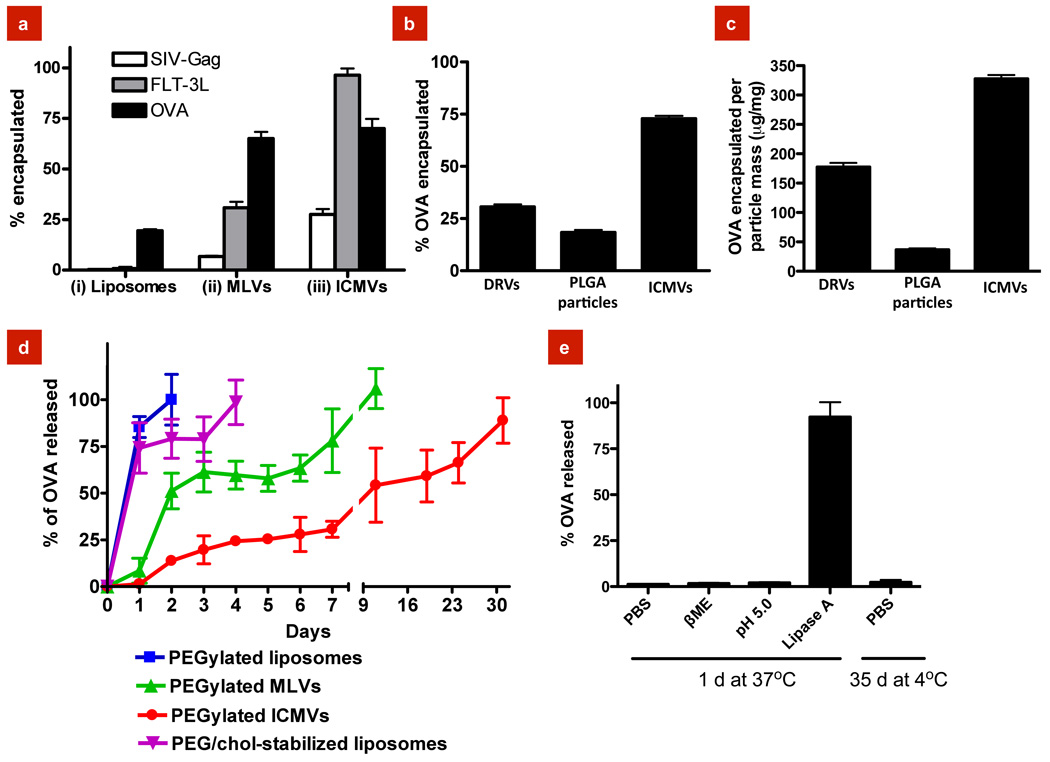
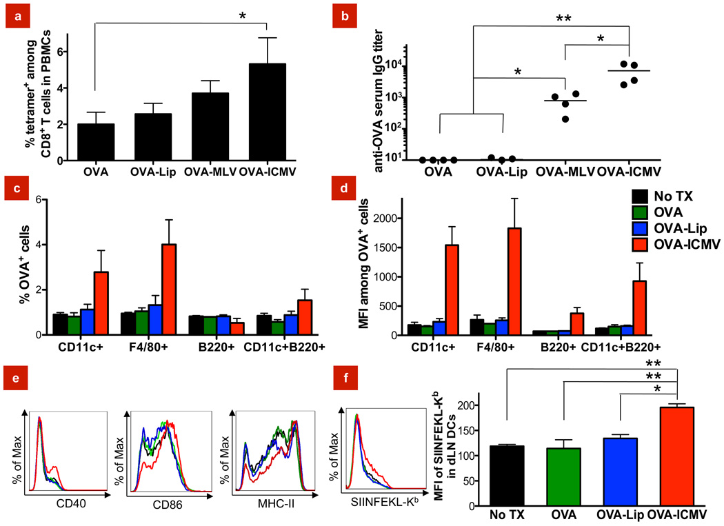
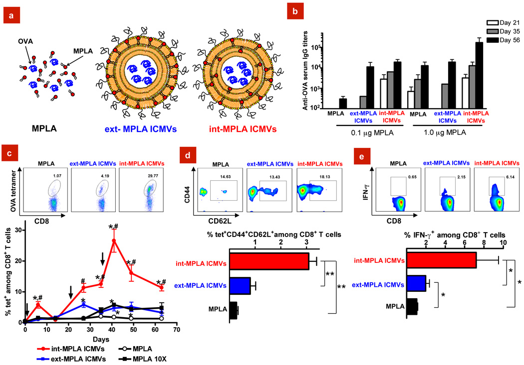
Vaccines based on recombinant proteins avoid the toxicity and antivector immunity associated with live vaccine (for example, viral) vectors, but their immunogenicity is poor, particularly for CD8(+) T-cell responses. Synthetic particles carrying antigens and adjuvant molecules have been developed to enhance subunit vaccines, but in general these materials have failed to elicit CD8(+) T-cell responses comparable to those for live vectors in preclinical animal models. Here, we describe interbilayer-crosslinked multilamellar vesicles formed by crosslinking headgroups of adjacent lipid bilayers within multilamellar vesicles. Interbilayer-crosslinked vesicles stably entrapped protein antigens in the vesicle core and lipid-based immunostimulatory molecules in the vesicle walls under extracellular conditions, but exhibited rapid release in the presence of endolysosomal lipases. We found that these antigen/adjuvant-carrying vesicles form an extremely potent whole-protein vaccine, eliciting endogenous T-cell and antibody responses comparable to those for the strongest vaccine vectors. These materials should enable a range of subunit vaccines and provide new possibilities for therapeutic protein delivery.


Figures

Figure 1
Figure 1. Synthesis of interbilayer-crosslinked multilamellar vesicles (ICMVs)
a, Schematic illustration of ICMV synthesis and cryoelectron microscope images: (i) Anionic, maleimide-functionalized liposomes are prepared from dried lipid films, (ii) divalent cations are added to induce fusion of liposomes and the formation of MLVs, (iii) membrane-permeable dithiols are added, which crosslink maleimide-lipids on apposed lipid bilayers in the vesicle walls, and (iv) the resulting lipid particles are PEGylated with thiol-terminated PEG. Cryo-EM images from each step of the synthesis show (i) initial liposomes, (ii) MLVs, and (iii) ICMVs with thick lipid walls. Scale bars = 100 nm. Right-hand image of (iii) shows a zoomed image of an ICMV wall, where stacked bilayers are resolved as electron-dense striations; scale bar = 20 nm. b, ICMV particle size histogram measured by dynamic light scattering. c, d, Histograms of ICMV properties from cryo-EM images show (c) the number of lipid bilayers per particle, and (d) the ratio of particle radius to lipid wall thickness. (n = 165 particles analyzed).
Figure 2
Figure 2. Protein encapsulation and release from ICMVs
a, Encapsulation efficiency of the globular proteins SIV-gag, FLT-3L, or OVA in lipid vesicles collected at each step of ICMV synthesis. b, c, Comparison of OVA encapsulation efficiency (b), and total protein loading per particle mass (c) in ICMVs vs. dehydration-rehydration vesicles (DRVs) or PLGA nanoparticles. d, Kinetics of OVA release from simple liposomes, MLVs, or ICMVs (all with base lipid composition 4:5:1 DOPC:MPB:DOPG) incubated in RPMI medium with 10% serum at 37°C measured over 30 days in vitro. Also shown for comparison are release kinetics for liposomes stabilized with cholesterol and PEG-lipid (38:57:5 DOPC:chol:PEG-DOPE). e, Release of OVA from ICMVs was measured in buffers simulating different aspects of the endolysomal environment: reducing buffer, 100 mM β-mercaptoethanol (β-ME) in PBS; acidic buffer, 50 mM sodium citrate pH 5.0; lipase-containing buffer, 500 ng/mL lipase A in PBS. Data represent the mean ± s.e.m of at least three experiments with n = 3.
Figure 3
Figure 3. In vitro stimulation of immune responses by ICMVs supplemented with the TLR agonist MPLA
a, Flow cytometry analysis of expression of the cell surface costimulatory markers CD40, CD80, and CD86 on splenic dendritic cells (DCs) after 18 hr incubation with 0.7 µg/mL soluble OVA, equivalent doses of OVA loaded in ICMVs, or ICMVs loaded with an irrelevant protein (vivax malaria protein, VMP), in the presence or absence of 0.1 µg/mL MPLA. b, Splenic DCs were incubated for 18 hr with 10 µg/mL SIINFEKL peptide (OVA257–264), 5.0 µg/mL soluble OVA, equivalent doses of OVA loaded in ICMVs, or VMP-loaded ICMVs in the presence or absence of 0.05 µg/mL MPLA, and the extent of cross-presentation of OVA was assessed by flow cytometry analysis of cells stained with the 25-D1.16 mAb that recognizes SIINFEKL complexed with H-2Kb. c, 5-(6)-carboxyfluorescein diacetate succinimidyl diester (CFSE)-labeled OVA-specific naïve OT-I CD8+ T-cells were co-cultured with syngeneic splenic DCs pulsed with soluble 0.7 µg/mL OVA mixed with 0.1 µg/mL MPLA, or equivalent doses of OVA-loaded ICMVs mixed with MPLA. Empty ICMVs without antigen or ICMVs loaded with the irrelevant antigen VMP were included as negative controls. Proliferation of CD8+ T-cells was assessed on day 3 by flow cytometry analysis of the dilution of CFSE in the OT-I CD8+ T-cells; shown are histograms of CFSE fluorescence. Gates on each histogram indicate the percentage of divided cells in each sample. Data represent the mean ± s.e.m of at least three experiments with n = 3–4.
Figure 4
Figure 4. In vivo immunization with ICMVs vs. soluble antigen or antigen encapsulated in non-crosslinked vesicles
a, b, C57Bl/6 mice were immunized s.c. with a single injection of 10 µg OVA delivered in soluble, liposomal, MLV, or ICMV formulations, each mixed with 0.1 µg of MPLA. a, The percentage of antigen-specific CD8+ T-cells was determined by flow cytometry analysis of peripheral blood mononuclear cells (PBMCs) 7 days post immunization with fluorescent OVA peptide-MHC tetramers. b, Sera from the immunized mice were analyzed by ELISA 21 days post immunization for OVA-specific IgG. c, d, C57Bl/6 mice were injected with 10 µg of fluorophore-conjugated OVA mixed with 0.1 µg of MPLA as a soluble, liposomal, or ICMV formulation, and the draining inguinal lymph node (dLN) cells that internalized OVA were assessed on day 2. c, Shown are percentages of DCs (CD11c+), macrophages (F4/80+), B cells (B220+), and plasmacytoid DCs (CD11c+B220+) positive for OVA uptake, and d, the mean fluorescence intensity (MFI) of OVA+ populations. e, f, C57Bl/6 mice were injected with 10 µg of OVA mixed with 0.1 µg of MPLA as a soluble, liposomal, or ICMV formulation, and 2 d later, DCs isolated from draining inguinal LNs were analyzed by flow cytometry to assess DC activation and antigen cross-presentation. e, Overlaid histograms show costimulatory markers (CD40 and CD86) and MHC-II expression in DCs. f, The left panel shows overlaid histograms of inguinal LN DCs stained for SIINFEKL-Kb+ complexes, and mean MFI levels are shown on the right panel. Data represent mean ± s.e.m of 2–3 independent experiments conducted with n = 3–4. *, p < 0.05 and **, p < 0.01, analyzed by one-way ANOVA, followed by Tukey’s HSD.
Figure 5
Figure 5. ICMVs carrying antigen in the aqueous core and MPLA embedded in the vesicle walls elicit potent antibody and CD8+ T-cell responses
a, Schematic illustration of the vaccine groups: soluble OVA mixed with MPLA (MPLA), OVA-loaded ICMVs with MPLA only on the external surface (ext-MPLA ICMVs), or OVA-loaded ICMVs with MPLA throughout the lipid multilayers (int-MPLA ICMVs). b–g, C57Bl/6 mice were immunized on days 0, 21, and 35 at tail base s.c. with 10 µg OVA and either 0.1 µg or 1.0 µg of MPLA formulated either as MPLA, ext-MPLA ICMVs, or int-MPLA ICMVs. b, ELISA analysis of total OVA-specific IgG in sera. c, Frequency of OVA-specific T-cells in peripheral blood assessed over time via flow cytometry analysis of tetramer+CD8+ T-cells for vaccinations with 10 µg OVA and 0.1 µg MPLA. Response to vaccinations with soluble OVA + 1 µg MPLA (MPLA 10X) also shown for comparison. Shown are representative flow cytometry scatter plots from individual mice at d41 and mean tetramer+ values from groups of mice vs. time. d, Analysis of T-cell effector/effector memory/central memory phenotypes in peripheral blood by CD44/CD62L staining on tetramer+ cells from peripheral blood on d41. Shown are representative cytometry plots from individual mice and mean percentages of tet+CD44+CD62L+ cells among CD8+ T-cells at d41. e, Functionality of antigen-specific CD8+ T-cells was assayed on d49 with intracellular IFN-γ staining after ex vivo restimulation of PBMCs with OVA peptide in vitro. Representative flow cytometry histograms of IFN-γ+CD8+ T-cells from individual mice and mean results from groups are shown. Data represent the mean ± s.e.m of two independent experiments conducted with n = 3. c, *, p < 0.05 compared to sol OVA + MPLA and #, p < 0.05 compared to ext-MPLA ICMVs. d, e, *, p < 0.05 and **, p < 0.01, analyzed by two-way ANOVA, followed by Tukey’s HSD.

Comment in

References

    1. Guy B. The perfect mix: recent progress in adjuvant research. Nat. Rev. Microbiol. 2007;5:505–517. - PubMed
    1. Perrie Y, Mohammed AR, Kirby DJ, McNeil SE, Bramwell VW. Vaccine adjuvant systems: enhancing the efficacy of sub-unit protein antigens. Int. J. Pharm. 2008;364:272–280. - PubMed
    1. Reed SG, Bertholet S, Coler RN, Friede M. New horizons in adjuvants for vaccine development. Trends Immunol. 2009;30:23–32. - PubMed
    1. Walker BD, Burton DR. Toward an AIDS vaccine. Science. 2008;320:760–764. - PubMed
    1. Haglund K, et al. Robust recall and long-term memory T-cell responses induced by prime-boost regimens with heterologous live viral vectors expressing human immunodeficiency virus type 1 Gag and Env proteins. J. Virol. 2002;76:7506–7517. - PMC - PubMed

Publication types