. 2011 Apr;6(4):253-60.
doi: 10.1038/nnano.2011.12. Epub 2011 Feb 20.

Controlling protein translocation through nanopores with bio-inspired fluid walls

Affiliations

Controlling protein translocation through nanopores with bio-inspired fluid walls

Erik C Yusko et al. Nat Nanotechnol. 2011 Apr.

Abstract

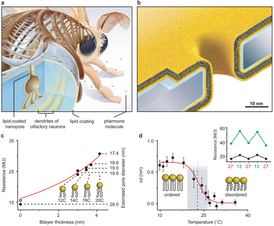
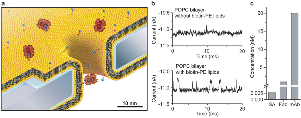
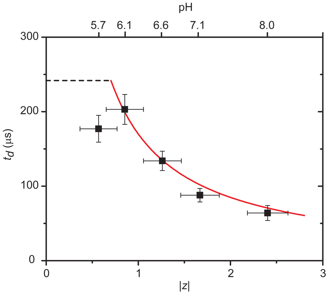
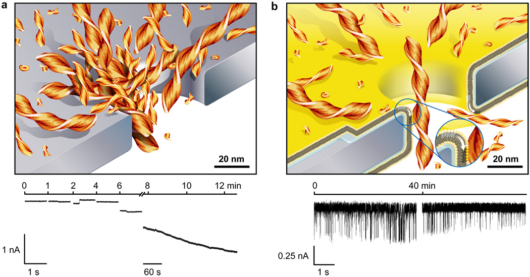
Synthetic nanopores have been used to study individual biomolecules in high throughput, but their performance as sensors does not match that of biological ion channels. Challenges include control of nanopore diameters and surface chemistry, modification of the translocation times of single-molecule analytes through nanopores, and prevention of non-specific interactions with pore walls. Here, inspired by the olfactory sensilla of insect antennae, we show that coating nanopores with a fluid lipid bilayer tailors their surface chemistry and allows fine-tuning and dynamic variation of pore diameters in subnanometre increments. Incorporation of mobile ligands in the lipid bilayer conferred specificity and slowed the translocation of targeted proteins sufficiently to time-resolve translocation events of individual proteins. Lipid coatings also prevented pores from clogging, eliminated non-specific binding and enabled the translocation of amyloid-beta (Aβ) oligomers and fibrils. Through combined analysis of their translocation time, volume, charge, shape and ligand affinity, different proteins were identified.


Conflict of interest statement

The authors declare no competing financial interests.

Figures

Figure 1
Figure 1. Bioinspired synthetic nanopores with bilayer-coated fluid walls
a, Cartoon showing a cross-section through one sensillum in the antenna of the silk moth Bombyx mori. Capture, pre-concentration, and translocation of pheromones through the exoskeleton of these sensilla towards dendrites of olfactory neurons is thought to occur via lipid-coated nanopores and pore tubules–. b, Cartoon, drawn to scale, showing a synthetic, lipid-coated (yellow) nanopore in a silicon nitride substrate (grey) and the interstitial water layer (blue). c, Nanopore resistance and corresponding open pore diameter as a function of the thickness of the bilayer coating. Red curve is a best fit of the data to equation (1). Numbers underneath the lipid cartoons refer to the number of carbons in their acyl chains (see Table 1). d, Actuation of nanopore diameters by a change in the thickness of the bilayer coating, Δd, in response to a thermal phase transition of DMPC lipids (see Supplementary Section S1). Blue dotted line and grey shaded region represent the mean value and range of phase transition temperatures reported for DMPC lipids. Inset: cycling the temperature between 13° and 27° C actuated the pore diameter dynamically as indicated by the larger changes in electrical resistance through a pore with (green squares) than without (back squares) a bilayer.
Figure 2
Figure 2. Capture, affinity-dependent pre-concentration, and translocation of specific proteins after binding to ligands on mobile lipid anchors
a, Cartoon, drawn to scale, illustrating binding of streptavidin (large red) to specific lipid-anchored biotin-PE (blue circles) followed by single molecule translocation of the anchored complex through the nanopore. b, Current versus time traces illustrating capture, pre-concentration, and reduced translocation speed of streptavidin. In the absence of biotin groups, only rare translocation events with short translocation times, td, could be detected in electrolytes containing 6 pM streptavidin (top current trace). In contrast, 0.4 mol% of biotinylated lipids in the lipid coating strongly increased the event frequency and slowed down the translocation speed sufficiently to enable complete time resolution of translocation events (bottom current trace). c, Minimum bulk concentrations of streptavidin, polyclonal anti-biotin Fab fragments, and monoclonal anti-biotin IgG antibodies required to observe at least 30 – 100 translocation events per second.
Figure 3
Figure 3. Controlling the translocation times, td, of single lipid-anchored proteins by the viscosity of the bilayer coating and distinguishing proteins by their most probable td values
a, Distribution of translocation times of streptavidin. Insets: current versus time traces illustrating that td could be prolonged more with intermediate viscosity POPC bilayers (blue current traces) than with low viscosity DΔPPC bilayers (red current traces). b, Translocation of anti-biotin Fab fragments through nanopores with bilayers of intermediate viscosity (POPC) or high viscosity (~49 mol% cholesterol and 50 mol% POPC). c, Translocation of anti-biotin antibodies through a pore with a coating of intermediate viscosity (POPC). Red, blue, and green curves represent a best fit of the corresponding data to a biased diffusion first passage time model (equation S10 in Supplementary Section S5). All bilayers contained 0.15 – 0.4 mol% biotin-PE. See Supplementary Sections S7 and S9 for binning methods, errors of td, and measurement errors.
Figure 4
Figure 4. Distribution of ΔI values and corresponding molecular volumes and shape factors of individual proteins translocating through bilayer-coated nanopores with biotinylated lipids
a–c, Translocation of streptavidin (a), anti-biotin Fab fragments (b) and anti-biotin antibodies (c); the dashed red lines indicate ΔI values that would be expected for IgG antibodies with a volume of 347 nm3 and different shape factors γ; see Supplementary Section S6 for a schematic illustration and discussion of shape factors,.
Figure 5
Figure 5. Comparison of experimental and theoretical values of charge-dependent translocation times of streptavidin
Experimental values are shown in black squares and the red curve represents the theoretical prediction by equation 3. Dashed black line corresponds to the expected translocation time for streptavidin assuming a translocation event due purely to diffusion in one dimension (td = <lp>2/(2DL), i.e. without an electrophoretic effect. The valance |z| of the net charge of streptavidin was varied by the pH of the electrolyte. The length of the pore with the bilayer coating was 28 ± 0.2 nm. Note that the red curve is not a best fit to the data; it is the prediction of td as a function of |z| according to equation (3) when all parameters were fixed to their known values.
Figure 6
Figure 6. Bilayer-coated nanopores resist clogging and enable the monitoring of the aggregation of amyloid-beta (Aβ) peptides
a, Cartoon illustrating clogging of uncoated nanopores and a typical current versus time trace during clogging of a nanopore by Aβ aggregates. This concatenated current trace shows several 1 s recordings and one 5 min recording. b, Cartoon illustrating translocation of individual Aβ aggregates through a bilayer-coated nanopore with a fluid wall (white arrow in the inset) and a typical current versus time trace of translocation events. The bilayer coating conferred non-fouling properties to these pores and enabled resistive pulse recordings over at least 40 min without clogging. Both recordings are 5 s long, one was taken immediately after addition of the Aβ sample and the other one 40 min later. Aβ (1–40) samples were aggregated for 72 h.

Comment in

References

    1. Sexton LT, et al. Resistive-pulse studies of proteins and protein/antibody complexes using a conical nanotube sensor. J. Am. Chem. Soc. 2007;129:13144–13152. - PubMed
    1. Movileanu L, Howorka S, Braha O, Bayley H. Detecting protein analytes that modulate transmembrane movement of a polymer chain within a single protein pore. Nature Biotech. 2000;18:1091–1095. - PubMed
    1. Howorka S, Siwy Z. Nanopore analytics: sensing of single molecules. Chem. Soc. Rev. 2009;38:2360–2384. - PubMed
    1. Siwy Z, et al. Protein biosensors based on biofunctionalized conical gold nanotubes. J. Am. Chem. Soc. 2005;127:5000–5001. - PubMed
    1. Ding S, Gao CL, Gu LQ. Capturing single molecules of immunoglobulin and ricin with an aptamer-encoded glass nanopore. Anal. Chem. 2009;81:6649–6655. - PMC - PubMed

Publication types