. 2011 Mar;13(3):223-33.
doi: 10.1038/ncb2165. Epub 2011 Feb 20.

How APC/C-Cdc20 changes its substrate specificity in mitosis

Affiliations

How APC/C-Cdc20 changes its substrate specificity in mitosis

Daisuke Izawa et al. Nat Cell Biol. 2011 Mar.

Erratum in

  • Nat Cell Biol. 2011 May;13(5):633

Abstract

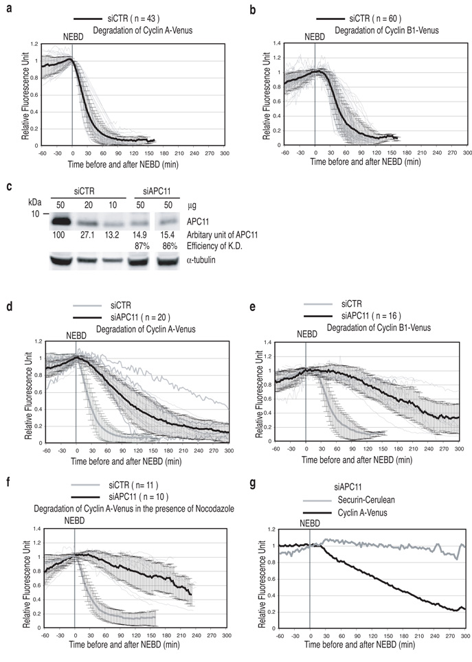
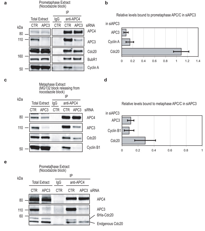
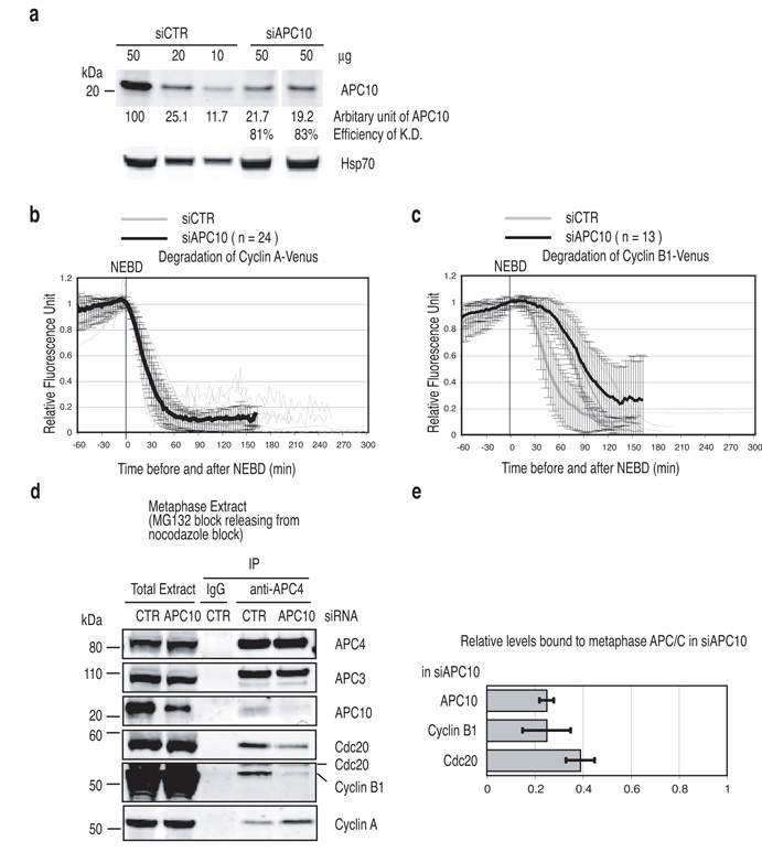
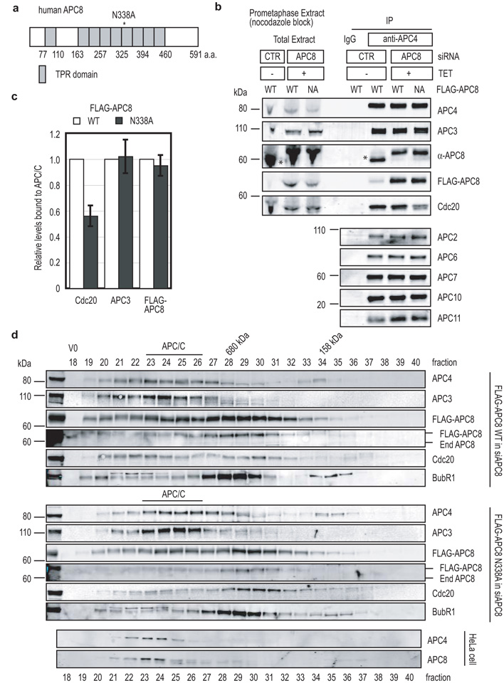
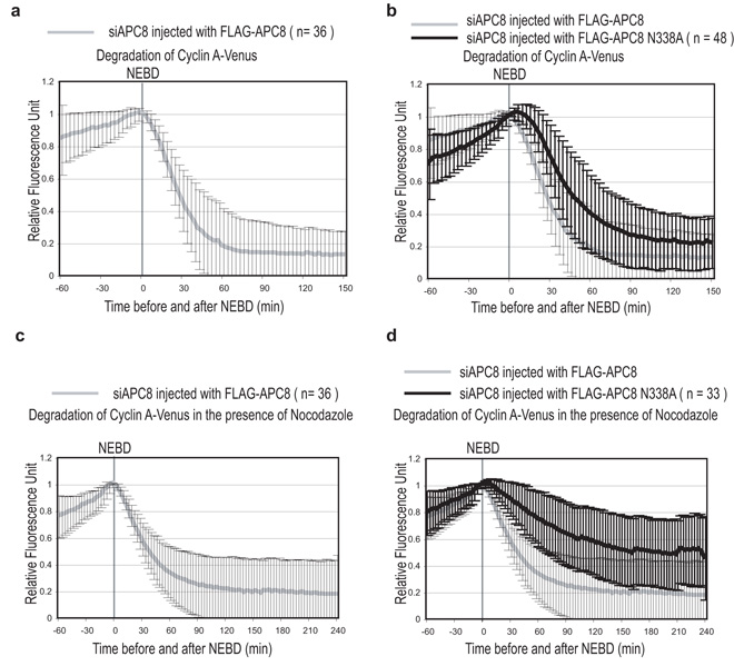
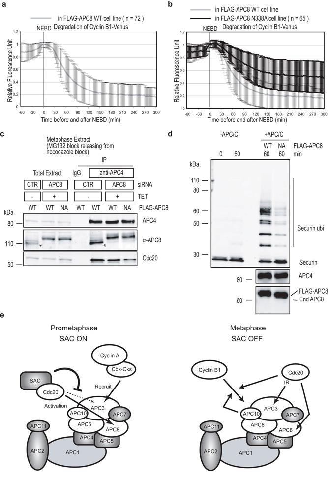
Progress through mitosis requires that the right protein be degraded at the right time. One ubiquitin ligase, the anaphase-promoting complex or cyclosome (APC/C) targets most of the crucial mitotic regulators by changing its substrate specificity throughout mitosis. The spindle assembly checkpoint (SAC) acts on the APC/C co-activator, Cdc20 (cell division cycle 20), to block the degradation of metaphase substrates (for example, cyclin B1 and securin), but not others (for example, cyclin A). How this is achieved is unclear. Here we show that Cdc20 binds to different sites on the APC/C depending on the SAC. Cdc20 requires APC3 and APC8 to bind and activate the APC/C when the SAC is satisfied, but requires only APC8 to bind the APC/C when the SAC is active. Moreover, APC10 is crucial for the destruction of cyclin B1 and securin, but not cyclin A. We conclude that the SAC causes Cdc20 to bind to different sites on the APC/C and this alters APC/C substrate specificity.


Figures

Figure 1
Figure 1
APC11 is required to degrade all model substrates (a and b) Degradation of model substrates in control cells. Cells were treated with siRNA oligos against GAPDH, injected with plasmids encoding Cyclin A-Venus (a) or Cyclin B1-Venus (b) in G2 phase and analysed by time-lapse DIC and fluorescence microscopy at 3 min intervals. The fluorescence of individual cells was measured, the value at NEBD set to 1 and plotted as thin light grey lines. The mean +/− s.d. for all cells is plotted as thick black lines. n = number of cells analyzed from 3 independent experiments. (c) Depletion of human APC11. Cells were treated with siRNA oligos against GAPDH (siCTR) or APC11 for 72 hrs before assaying the indicated amounts of extract by quantitative immunoblotting. The extent of depletion was calculated from a standard curve using diluted control extract and normalisation to the level of tubulin. Results are representative of at least two experiments per siRNA treatment. (d and e) APC11 is required to degrade both SAC–sensitive and SAC-insensitive substrates. Cells were treated with siRNA oligos against APC11, injected with a plasmid encoding Cyclin A-Venus (d) or Cyclin B1-Venus (e) and analyzed as in a. The mean +/− s.d. for all cells are plotted as the thick black lines. The experimental values are plotted in black (siAPC11) and the controls are plotted in grey (siCTR). n = number of cells analyzed from 3 independent experiments. (f) The SAC stabilises Cyclin A-Venus in cells depleted of APC11. Cells were treated with siRNA oligos against GAPDH (grey) or APC11 (black), injected with a plasmid expressing Cyclin A-Venus, treated with 100 ng-ml nocodazole and analysed as in a. n = number of cells analyzed from 2 independent experiments. (g) Cyclin A is preferred over securin as a substrate when APC-C activity is limiting. Cells were treated with siRNA oligos against APC11, injected with a plasmid expressing Cyclin A-Venus (black) and securin-Cerulean (grey) and analyzed as in a. Representative of 11 cells in 2 independent experiments.
Figure 2
Figure 2
APC3 is required to degrade both SAC-sensitive and SAC–resistant substrates. (a) Depletion of human APC3. Cells were treated with siRNA oligos against GAPDH or APC3 for 72 hrs before analyzing. Protein levels were analyzed as in Fig 1c. (b and c) APC3 is required to degrade both SAC–insensitive and SAC-sensitive substrates. Cells were treated with siRNA oligos against APC3, injected with a plasmid encoding Cyclin A-Venus (b) or Cyclin B1-Venus (c) and analyzed as in Fig 1a. The mean +/− s.d. is plotted as thick black lines. The experimental values are plotted in black (siAPC3) and the values from the control GAPDH-depleted cells are plotted in grey (siCTR). n = number of cells analyzed from 3 independent experiments. (d) APC-C composition in the depletion of APC3 or APC10. HeLa cells were treated with siRNA against GAPDH, APC3 or APC10 for 72 hrs before harvesting prometaphase cells in the presence of 100 ng-ml nocodazole by mitotic shake off. The APC-C was immunoprecipitated using anti-APC4 antibodies and the immunoprecipitates blotted with antibodies against phospho-APC1 (serine 355), APC2, APC3, APC4, APC6, APC7, APC8, APC10 and APC11, and the extent of depletion measured by quantitative immunoblotting for the cell extracts (top) and immunoprecipitates (bottom). (e) Analysis of APC-C in the depletion of APC3 by size-exclusion chromatography. Extracts of control (top) or APC3-depleted (bottom) cells were separated on a Superose 6 column and fractions were blotted with antibodies against APC3, APC4 and APC6. The total cell extract is also shown (top right). Loading control refers to a cross-reacting protein recognised by the anti-APC3 antibody. The peak of APC-C migration is indicated by the black bar.
Figure 3
Figure 3
APC3 is only required to bind Cdc20 when the SAC is satisfied (a - d) APC3 is only required to bind Cdc20 when the SAC is satisfied. HeLa cells were treated with siRNA against GAPDH (CTR) or APC3 and synchronised in prometaphase by treating with 100 ng-ml nocodazole plus 10 μM MG132 to stabilise Cyclin A (a & b), or synchronised in prometaphase with 100 ng-ml nocodazole, released into medium containing 10 μM MG132 and incubated for a further 3 hrs to obtain metaphase cells (c & d). The APC-C complex was immunoprecipitated with anti-APC4 antibodies and samples blotted for APC3, APC4, Cdc20, BubR1 and Cyclin A (a) or Cyclin B1 (b). (b and d) Bar diagrams show the remaining amount of APC3 and the amount of Cdc20 and Cyclin A or B1 bound to the prometaphase APC-C (b) or the metaphase APC-C (d), quantified using a LI-COR Odyssey scanner and normalised to the level of APC4. Levels of the proteins bound to control APC-C were set to 1. Error bars shown are mean+/− s.d. of 3 experiments. (e) APC3 is required for free Cdc20 to bind to the APC-C. Cells were treated with siRNA oligos against GAPDH or APC3 and synchronized as in (a). Purified recombinant His6-tagged Cdc20 was added to the cell extracts and the APC-C was immunoprecipitated with an anti-APC4 antibody. Samples were blotted for APC3, APC4 and Cdc20. Recombinant Cdc20 runs at a higher molecular mass than endogenous Cdc20.
Figure 4
Figure 4
APC10 is required to recruit SAC-sensitive substrates (a) Depletion of APC10. Cells were treated with siRNA oligos against GAPDH (control) or APC10 and the results from 2 independent experiments analyzed as in Fig 1c. Hsp70 was used as a loading control. (b and c) APC10 is required for Cyclin B1 but not for Cyclin A degradation. Cells were treated with siRNA oligos against APC10, injected with a plasmid encoding Cyclin A-Venus (b) or Cyclin B1-Venus (c) and analysed by time-lapse as Fig 1a. The mean +-− s.d. for all cells is plotted in black (siAPC10) and the values from the control cells are plotted in grey (siCTR). n = number of cells analyzed from 2 independent experiments. (d and e) APC10 mediates Cyclin B1 and Cdc20 but not Cyclin A binding to APC-C. HeLa cells were treated with siRNA against GAPDH (CTR) or APC10 and synchronised in metaphase as in Fig 3c. The APC-C complex was immunoprecipitated with anti-APC4 antibodies and samples blotted for APC3, APC4, APC10, Cdc20, Cyclin A and Cyclin B1 (d). (e) Bar diagrams show the remaining amount of APC10 and the amount of Cyclin B1 and Cdc20 bound to the metaphase APC-C quantified using a LI-COR Odyssey scanner and normalised to the level of APC4. Levels of the proteins bound to control APC-C were set to 1. The mean +/− s.d. of 3 experiments is shown.
Figure 5
Figure 5
A point mutation in APC8 is sufficient to reduce the binding of Cdc20 in prometaphase (a) Schematic structure of human APC8. TPR domains are shown as grey boxes. (b) The N338A mutation reduces Cdc20 binding to prometaphase APC-C. HeLa cells with an inducible wild type Flag-APC8 (WT) or mutant Flag-APC8N338A were treated with siRNA against GAPDH (CTR) or APC8 and synchronised in prometaphase as in Fig 2d. The APC-C was immunoprecipitated using anti-APC4 antibodies and the immunoprecipitates blotted with antibodies against APC2, APC3, APC4, APC6, APC7, APC8, APC10, APC11 and Cdc20 and the extent of depletion measured by quantitative immunoblotting. The asterisk indicates endogenous APC8. (c) Quantification of APC3, Cdc20 and Flag-APC8 bound to the APC-C by quantitative immunoblotting and normalised to the level of APC4. Levels of the proteins bound to control APC-C were set to 1. The mean +/− s.d. of 4 independent experiments is shown. (d) Analysis of APC-C by size-exclusion chromatography. Control (bottom) and experimental HeLa cells expressing an inducible wild type Flag-APC8 (WT, top) or mutant Flag-APC8N338A (middle) were treated with siRNA oligos against APC8 and synchronised in prometaphase as in panel b and extracts prepared as in Fig. 3e. Cell extracts were fractioned on a Superose 6 column and fractions were blotted with antibodies against APC3, APC4, Flag epitope, APC8, Cdc20 and BubR1. The peak of APC-C migration is indicated by the black bar.
Figure 6
Figure 6
A point mutation in APC8 prevents Cyclin A degradation in prometaphase (a and b) Cyclin A degradation is delayed in cells expressing APC8N338A. Cells were treated with siRNA oligos against APC8, injected with a plasmid encoding Cyclin A-Venus and Flag-APC8 WT (a) or Flag-APC8N338A (b) and analysed by time-lapse DIC and fluorescence microscopy as in Fig 1a. The experimental mean +/− s.d. of wild type APC8 is plotted in grey in panel a and b, and of APC8 N338A plotted in black. n = number of cells analyzed in 3 independent experiments. (c and d) Cyclin A is stabilized by nocodazole in cells expressing APC8N338A. Cells were treated with siRNA oligos against APC8, injected with a plasmid encoding Cyclin A-Venus and wild type Flag-APC8 (c) or Flag-APC8N338A (d) in G2 phase and analysed in the presence of 100 ng-ml nocodazole by time-lapse DIC and fluorescence microscopy. The experimental mean +/− s.d. of wild type APC8 is plotted in grey in panel c and d, and of APC8N338A plotted in black. n = number of cells analyzed from 3 independent experiments.
Figure 7
Figure 7
The Cdc20-binding site on APC8 is also required in metaphase (a and b) Cyclin B1 degradation is inhibited in cells expressing APC8N338A. siRNA against APC8 and a plasmid encoding Cyclin B1-Venus were transfected into the HeLa cells with an inducible siRNA-resistant wild type Flag-APC8 (a) or mutant Flag-APC8N338A (b) and analysed as in Fig 1a. Mean +/− s.d. values are shown. n = number of cells analyzed from 3 independent experiments. (c) APC8 is important for free-Cdc20 to bind to the APC-C. HeLa cells expressing an inducible wild type Flag-APC8 (WT) or mutant Flag-APC8N338A were treated with siRNA against GAPDH (CTR) or APC8 as in Fig 5b and synchronised in metaphase as in Fig 3c. The APC-C was immunoprecipitated using anti-APC4 antibodies and the immunoprecipitates blotted with antibodies against APC4, APC8 and Cdc20. The asterisk indicates endogenous APC8. (d) APC8 N338A mutation reduces APC-C activity in vitro. The APC-C incorporating Flag-APC8 or Flag-APC8N338A was prepared as in Fig 5b and its activity assayed using securin as a substrate in an in vitro ubiquitination reaction as previously described . (e) Model for APC-C regulation during early mitosis. In prometaphase (left), the APC-C recognizes substrates such as Cyclin A through the APC3 subunit but interacts through APC8 with Cdc20, as co-activator or as part of the SAC complex. The SAC proteins associated with Cdc20 could prevent Cdc20 accessing its APC3 binding site. In metaphase (right), the APC-C recognizes substrates such as Cyclin B1 through APC10 and Cdc20 forming a bi-partite receptor, and Cdc20 requires both APC3 and APC8 to interact with and activate the APC-C.

Similar articles

Cited by

References

    1. Pines J. Mitosis: a matter of getting rid of the right protein at the right time. Trends Cell Biol. 2006;16:55–63. - PubMed
    1. Peters JM. The anaphase promoting complex-cyclosome: a machine designed to destroy. Nature reviews. 2006;7:644–656. - PubMed
    1. Thornton BR, et al. An architectural map of the anaphase-promoting complex. Genes & development. 2006;20:449–460. - PMC - PubMed
    1. Gmachl M, Gieffers C, Podtelejnikov AV, Mann M, Peters JM. The RING-H2 finger protein APC11 and the E2 enzyme UBC4 are sufficient to ubiquitinate substrates of the anaphase-promoting complex. Proceedings of the National Academy of Sciences of the United States of America. 2000;97:8973–8978. - PMC - PubMed
    1. Tang Z, et al. APC2 Cullin protein and APC11 RING protein comprise the minimal ubiquitin ligase module of the anaphase-promoting complex. Molecular biology of the cell. 2001;12:3839–3851. - PMC - PubMed

Publication types