Comparative Study
. 2011 Sep;20(9):1434-40.
doi: 10.1007/s00586-011-1720-y. Epub 2011 Feb 19.

Comparison between pedicle subtraction osteotomy and anterior corpectomy and plating for correcting post-traumatic kyphosis: a multicenter study

Affiliations
Comparative Study

Comparison between pedicle subtraction osteotomy and anterior corpectomy and plating for correcting post-traumatic kyphosis: a multicenter study

Mohammad M El-Sharkawi et al. Eur Spine J. 2011 Sep.

Abstract

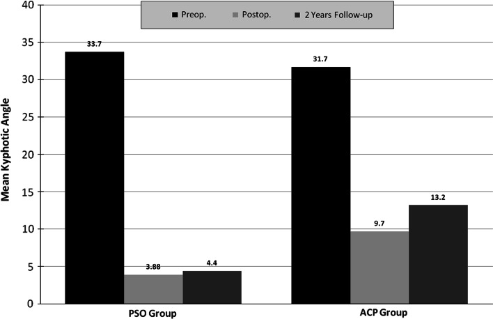
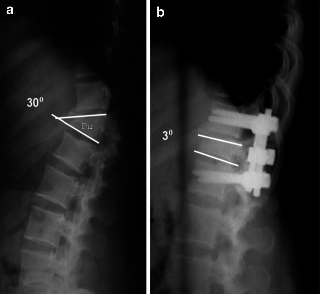
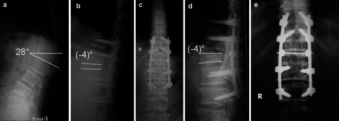
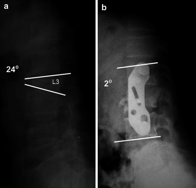
Kyphosis is a common sequel of inadequately managed thoracolumbar fractures. This study compares between pedicle subtraction osteotomy (PSO) and anterior corpectomy and plating (ACP) for correcting post-traumatic kyphosis. Forty-three patients with symptomatic post-traumatic kyphosis of the thoracolumbar spine were treated with PSO and prospectively followed for a minimum of 2 years. Visual Analogue Scale (VAS) and Oswestry Disability Index (ODI) were used to assess clinical improvement and radiographs were obtained at 2, 6, 12 and 24 months. The recorded clinical and radiological outcomes were compared to a control group of 37 patients, who were treated earlier by the same authors with ACP. The mean correction of the kyphotic angle was 29.8° for the PSO group and 22° for the ACP group (P = 0.001). PSO group showed significantly better improvement in the VAS score and the ODI. At final follow-up, patients reported very good satisfaction (93% in PSO vs. 81% in ACP) and good function (90% in PSO vs. 73% in ACP). Complications in the PSO group included pulling out of screws and recurrence of deformity requiring revision and longer fixation (1 patient), and transient lower limb paraesthesia (2 patients). Recorded complications in the ACP group included an aortic injury (1 patient) that was successfully repaired, pseudarthrosis (1 patient), persistent graft donor site morbidity (3 patients), and incisional hernia (1 patient). PSO and ACP are demanding procedures. PSO seems to be equally safe but more effective than ACP for correcting post-traumatic kyphosis.


Figures

Fig. 1
Fig. 1
Changes in mean kyphotic angle in the PSO and ACP groups
Fig. 2
Fig. 2
a Lateral X-ray of a 41-year-old manual worker who is suffering from severe back pain and inability to work for 1 year due to post-traumatic kyphosis of D12 of 30°. b Two years follow-up lateral X-ray after PSO shows excellent correction of the deformity to 3°. This patient returned to his previous work 9 months postoperatively
Fig. 3
Fig. 3
A 27-year-old accountant presented with severe back pain and deformity and inability to work. Five months earlier, he had percutaneous fixation of his L1 fracture in another hospital. a Lateral X-ray shows post-traumatic kyphosis of L1 of 28°. b Two years follow-up X-ray after PSO shows excellent correction of the deformity to (−4)°. He resumed his previous work 6 months postoperatively
Fig. 4
Fig. 4
a Lateral X-ray of a 58-year-old worker who is suffering from severe back pain and inability to work for 15 months due to post-traumatic kyphosis of L1 of 28°. b, c Postoperative X-rays show excellent correction of the deformity to (−4°). d, e Two years follow-up X-rays after PSO show maintenance of deformity correction and solid fusion. He resumed his previous work 6 months postoperatively. Although the distal screws seem to have pulled out partially, this was asymptomatic and did not affect the degree of local kyphosis correction. This backing out of the very distal screws was observed in the 2-month radiographs and was attributed to overcontouring of the rods
Fig. 5
Fig. 5
a Lateral X-ray of a 33-year-old housewife who is suffering from severe back pain for the last 7 months due to post-traumatic kyphosis of L3 of 24°. b Two years follow-up lateral X-ray following ACP shows significant correction of her deformity to 2°. She fully resumed her activities of daily living 4 months postoperatively, despite the suboptimal correction of her deformity

References

    1. Roberson JR, Whitesides TE. Surgical reconstruction of late post-traumatic thoracolumbar kyphosis. Spine. 1985;10:307–312. doi: 10.1097/00007632-198505000-00003. - DOI - PubMed
    1. Gertzbein SD, Harris MB. Wedge osteotomy for the correction of posttraumatic kyphosis: a new technique and a report of three cases. Spine. 1992;17:374–379. doi: 10.1097/00007632-199203000-00025. - DOI - PubMed
    1. Malcolm BW, Bradford DS, Winter RB, Chou SN. Post-traumatic kyphosis: a review of forty-eight surgically treated patients. J Bone Joint Surg Am. 1981;63:891–899. - PubMed
    1. Kostuik JP, Matsusaki H. Anterior stabilization instrumentation, and decompression for post-traumatic kyphosis. Spine. 1989;14:379–386. doi: 10.1097/00007632-198904000-00006. - DOI - PubMed
    1. Oda I, Cunningham BW, Buckley RA, Goebel MJ, Haggerty CJ, Orbegoso CM, McAfee PC. Does spinal kyphotic deformity influence the biomechanical characteristics of the adjacent motion segments? An in vivo animal model. Spine. 1999;24:2139–2146. doi: 10.1097/00007632-199910150-00014. - DOI - PubMed

MeSH terms