

Relationship between epicardial fat measured by 64-multidetector computed tomography and coronary artery disease
- PMID: 21337349
- PMCID: PMC6652334
- DOI: 10.1002/clc.20840
Relationship between epicardial fat measured by 64-multidetector computed tomography and coronary artery disease
Abstract
Background: Epicardial fat (EF) is the visceral fat of the heart deposited under the visceral layer of the pericardium and has the same origin as abdominal visceral fat, which is shown to be strongly related to the development of coronary artery disease (CAD). We measured the volume of EF (EFV) by 64-multidetector computed tomography (MDCT) and studied the relationship between EFV and the severity of CAD.
Hypothesis: Epicardial fat volume increases steeply in patients with significant coronary artery stenosis and in those with severe coronary artery calcification.
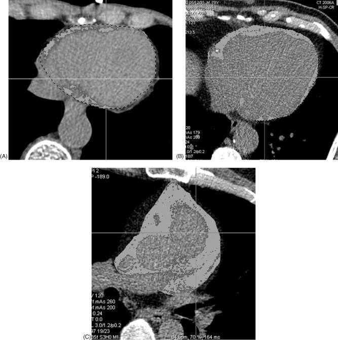
Methods: We studied 197 patients with suspected CAD who underwent 64-MDCT and coronary angiography. Cross-sectional tomographic cardiac slices (3.0 mm thick) from base to apex (30 to 40 slices per heart) were traced semiautomatically and EFV was measured by assigning Hounsfield units ranging from -30 to -250 to fat.
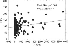
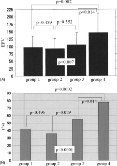
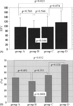
Results: Epicardial fat volume was 99.4 ± 40.0 ml (range, 11.6 to 263.8 mL) and coronary artery calcium score (CACS) was 267.2 ± 605.1 (range, 0 to 3780). There was a significant relationship between EFV and CACS (r=0.210, P=0.003). Patients with EFV >100 had a CACS that was significantly higher than in those with EFV <100 (384.0 ± 782.0 vs 174.8 ± 395.6; P = 0.016). The incidence of significant CAD was significantly higher in patients with EFV >100 compared with those with EFV <100 (40.2% vs 22.7%; P=0.008). The EFV was significantly higher in patients with severe coronary artery stenosis and in those with severe coronary artery calcification (CACS >400).
Conclusions: Our results showed that EFV was associated with coronary atherosclerosis, and EFV increased steeply in patients with severe coronary artery stenosis and in those with severe coronary artery calcification.
© 2011 Wiley Periodicals, Inc.
Figures
Comment in
-
Epicardial adipose tissue: an emerging role for the development of coronary atherosclerosis.Clin Cardiol. 2011 Mar;34(3):143-4. doi: 10.1002/clc.20893. Clin Cardiol. 2011. PMID: 21400540 Free PMC article.
References
-
- Mazurek T, Zhang L, Zalewsky A, et al. Human epicardial adipose tissue is a source of inflammatory mediators. Circulation. 2003;108:2460–2466. - PubMed
-
- Iacobellis G, Pistilli D, Gucciardo M, et al. Adiponectin expression in human epicardial adipose tissue in vivo is lower in patients with coronary artery disease. Cytokine. 2005;29:251–255. - PubMed
-
- Chaowalit N, Somers VK, Pellikka PA, et al. Subepicardial adipose tissue and the presence and severity of coronary artery disease. Atherosclerosis. 2006;186:354–359. - PubMed
-
- Jeong JW, Jeong MH, Yun KH, et al. Echocardiographic epicardial fat thickness and coronary artery disease. Circ J. 2007;71: 536–539. - PubMed
MeSH terms
LinkOut - more resources
Full Text Sources
Medical
Miscellaneous
