. 2011 Mar;240(3):577-88.
doi: 10.1002/dvdy.22477. Epub 2010 Nov 8.

MESD is essential for apical localization of megalin/LRP2 in the visceral endoderm

MESD is essential for apical localization of megalin/LRP2 in the visceral endoderm

Janet K Lighthouse et al. Dev Dyn. 2011 Mar.

Abstract

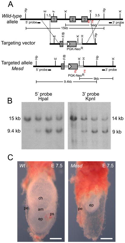
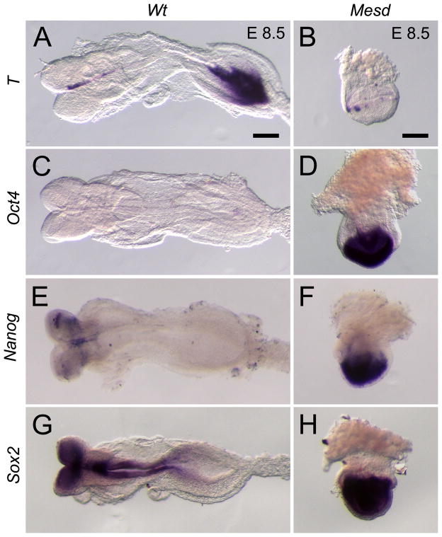
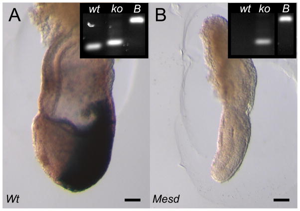
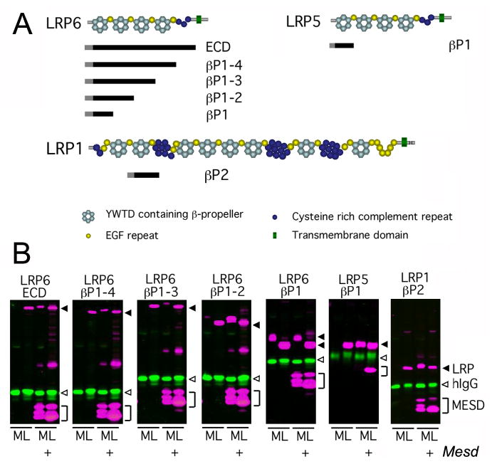
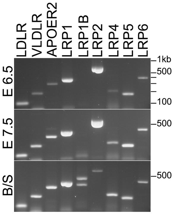
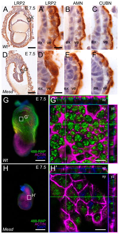
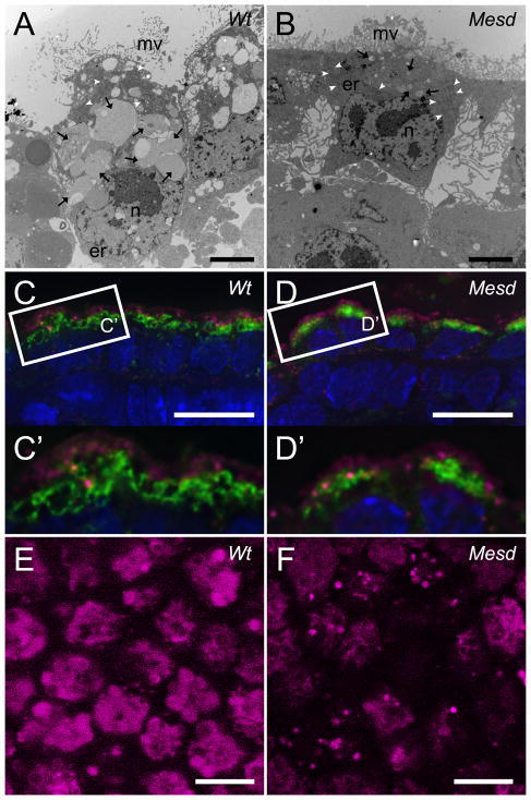
Deletion of the Mesd gene region blocks gastrulation and mesoderm differentiation in mice. MESD is a chaperone for the Wnt co-receptors: low-density lipoprotein receptor-related protein (LRP) 5 and 6 (LRP5/6). We hypothesized that loss of Wnt signaling is responsible for the polarity defects observed in Mesd-deficient embryos. However, because the Mesd-deficient embryo is considerably smaller than Lrp5/6 or Wnt3 mutants, we predicted that MESD function extends more broadly to the LRP family of receptors. Consistent with this prediction, we demonstrated that MESD function in vitro was essential for maturation of the β-propeller/EGF domain common to LRPs. To begin to understand the role of MESD in LRP maturation in vivo, we generated a targeted Mesd knockout and verified that loss of Mesd blocks WNT signaling in vivo. Mesd mutants continue to express the pluripotency markers Oct4, Nanog, and Sox2, suggesting that Wnt signaling is essential for differentiation of the epiblast. Moreover, we demonstrated that MESD was essential for the apical localization of the related LRP2 (Megalin/MEG) in the visceral endoderm, resulting in impaired endocytic function. Combined, our results provide evidence that MESD functions as a general LRP chaperone and suggest that the Mesd phenotype results from both signaling and endocytic defects resulting from misfolding of multiple LRP receptors.


Figures

Figure 1
Figure 1. Generation of Mesdtm1bch (Mesd-KO)
(A) Targeted disruption of Mesd. The coding region of Mesd contains three exons (numbered gray boxes). In the targeting vector the neomycin resistance cassette partially replaces exon 1 and the entirety of exons 2 and 3. Regions of homology are indicated with dashed lines. Genotyping primer pairs are shown as red bars and labeled in red as a, b, or c. (B) Southern blot of ES cell clones using a 5′ probe with HpaI digestion and a 3′ probe with KpnI digestion indicated successful disruption of Mesd. (C) The general phenotype of the Mesd-KO mutant compared to a wild-type littermate at E 7.5 was similar to the phenotype of the Mesd deletion phenotype previously observed (Holdener et al., 1994; Wines et al., 2000). ps, primitive streak; pe, parietal endoderm; ep, epiblast; am, amnion; ch, chorion. Scale bars in C indicate 500μm.
Figure 2
Figure 2. Expression of pluripotency markers persists in E 8.5 Mesd-KO embryos
(A) By E 8.5, wild-type embryos express T in the notochord and tail mesenchyme and have down-regulated or regionalized expression of (C) Oct4, (E) Nanog, and (G) Sox2 in the epiblast. In contrast, in E 8.5 Mesd-KO littermates, expression of (B) T in the extra-embryonic ectoderm and (D) Oct4, (F) Nanog, and (H) Sox2 in the epiblast was more consistent with that typically observed in a pre-gastrula embryo (Scholer et al., 1990; Wilkinson et al., 1990; Avilion et al., 2003; Chambers et al., 2003; Hart et al., 2004; Rivera-Perez and Magnuson, 2005), suggesting that Mesd-KO embryos were considerably delayed in development and that differentiation was blocked. Scale bars indicate 100μm. E 8.5 mutant embryos are oriented so the anterior-posterior axis is in the z-plane.
Figure 3
Figure 3. WNT signaling is blocked in E 7.5 Mesd-KO embryos
(A) At E 7.5, embryos heterozygous for Mesd-KO activated the BAT-gal reporter in the primitive streak and migrating mesoderm. (B) Although Mesd deficient embryos express Wnt3 at E7.5 (Hsieh et al., 2003), Mesd-KO littermates did not form primitive streak or mesoderm and did not activate BAT-gal reporter, indicating a defect in WNT signaling. (Insets) PCR genotyping confirmed embryo genotype. wt, wild-type allele; ko, Mesd-KO allele; B, BAT-gal allele. Scale bars indicate 100 μm. E 7.5 embryos are oriented with the anterior-posterior axis on the x-y plane, with the anterior on the left and posterior on the right.
Figure 4
Figure 4. MESD promotes trafficking of β-propeller/EGF domains
We utilized a soluble receptor secretion assay to identify the minimal LRP domain that requires MESD for maturation. In this assay, soluble truncated receptors were co-transfected with or without Mesd, and the cell lysate (L) and media (M) collected. If the soluble receptor is dependent upon Mesd for maturation, the receptor is expected to accumulate in the cell lysate (L) in the absence of exogenous Mesd. In the presence of exogenous Mesd, the soluble receptor would transit the secretory pathway and be released in to the cell culture media (M). (A) Overview of soluble receptor constructs. Pictured above is a schematic representation of the predicted domain structure of the full length LRP6, LRP5, and LRP1 receptors. The extra-cellular domain (ECD) of LRPs consists of cysteine-rich complement-like repeats (CLRs, dark blue circles), Epidermal growth factor (EGF) repeats (yellow circles), and alternating YWTD containing β-propeller (light blue hexagon) and EGF domains (LRP cartoons adapted from (Strickland et al., 2002)). The black bars located below the full length LRPs indicate the portion of receptor retained in the soluble receptor constructs. The receptor construct name, indicating the β-propeller/EGF domains (βP) included in the construct, is designated to the right of the black bar. Note β-propeller/EGFs are numbered sequentially starting at the N-terminus. All soluble receptors lacked the transmembrane domain (green triangle) present in the full length LRPs, but retained the signal peptide (grey bar) and maintain the juxtaposition of the β-propeller and C-terminal EGF motif (B) Western analysis of soluble LRP5/6 or LRP1 constructs in the presence or absence of MESD. Secreted receptors were detected in the cell culture media (M), and immature receptors were detected in the cell lysate (L). Closed arrowhead, LRP (magenta); open arrowhead, control hIgG (green); bracket, MESD (magenta).
Figure 5
Figure 5. LRPs containing one or more β-propeller/EGF domains are expressed in wild-type embryos at E 6.5 and E 7.5
Expression of LRP family members at E 6.5 (top) and E 7.5 (middle). LRP1B was the only LRP member not expressed at E 6.5 and 7.5. Primers detected both LRP1B isoforms in cDNA from brain and spleen (bottom).
Figure 6
Figure 6. LRP2 requires MESD for apical membrane localization at E 7.5
Immunohistochemistry indicated LRP2, CUBN, and AMN were apically localized in wild-type VE (A, A′, B, and C). In contrast, LRP2 was distributed diffusely throughout the Mesd-KO VE (D, D′), consistent with ER retention of improperly folded receptor (Hsieh et al.). In contrast, apical localization of CUBN and AMN was not affected by loss of MESD (E, F). Comparison of visceral endoderm endocytosis of 488-RAP in E 7.5 wild-type (G, G′) and Mesd mutant littermates (H, H′) indicated a reduction in functional LRP in Mesd VE. Black boxes in A and D indicate the region magnified in A′, B, C and D′, E, and F. White boxes in G and H indicate the region shown at in G′ and H′. Scale bars in A, D, G, and H indicate 100μm, and in A′, D′, G′, and H′ indicate 10μm. Note that G′ and H′ are shown at the same magnification; however, because orthogonal planes shown in G′ and H′ were chosen to maximize signal from endocytosed 488-RAP, some cells in H′ (or Supplement Figure 2B) appear larger than 488-RAP labeled wild-type due to imaging at a different z-planes. ex – extraembryonic ectoderm, ve – visceral endoderm.
Figure 7
Figure 7. Lysosome size is reduced in Mesd-KO VE at E 7.5
(A, B) TEM of sectioned embryonic VE from E 7.5 littermates. (A) Wild-type VE contained numerous small and large membrane-bound compartments (black arrows) concentrated apically relative to the nucleus (n). Wild-type VE cells contacted neighbors along the apical/basal boundary and thin microvilli (mv) extend from the apical surface. (B) VE of Mesd-KO embryos appeared shorter due to a decrease in vesicle size. Mutant VE had small membrane-bound compartments (black arrows), and the basal surface of the VE appeared to have lost contact with neighboring cells and the basement membrane. The apical surface had shorter and thicker microvilli (mv). TEM images taken at 4200x. Scale bars indicate 2 μm. er – endoplasmic reticulum, white arrowheads – mitochondria. (C) Identification of early endosomes (EEA1, magenta) and lysosomes (LAMP1/2, green) and nuclei (DAPI, blue) in wild-type VE at E 7.5. Early endosomes were more apical than the large, ring-like lysosomal structures. (D) We observed a reduction in the size of LAMP-positive bodies in Mesd VE at E 7.5, without a noticeable reduction in EEA1-positive bodies. (C′ and D′) Magnification of boxed areas in (C) and (D). (E) Staining of wild-type VE at E 7.5 with 10nM of LysoTracker Red revealed large, round lysosomes at least 2μm in diameter. (F) Staining of Mesd VE at E 7.5 showed smaller and fewer lysosomes, between 1μm and 2μm in diameter. Images were deconvolved using the Zeiss iterative deconvolution filter and nine images through 1.8μm were stacked to produce the images shown in Figure C and D. A single deconvolved Z-stack is shown in Figure C′ and D′. Scale bars in (C, D, E, F) indicate 10μm.

References

    1. Andersen OM, Christensen LL, Christensen PA, Sorensen ES, Jacobsen C, Moestrup SK, Etzerodt M, Thogersen HC. Identification of the minimal functional unit in the low density lipoprotein receptor-related protein for binding the receptor-associated protein (RAP). A conserved acidic residue in the complement-type repeats is important for recognition of RAP. J Biol Chem. 2000;275:21017–21024. - PubMed
    1. Artus J, Panthier JJ, Hadjantonakis AK. A role for PDGF signaling in expansion of the extra-embryonic endoderm lineage of the mouse blastocyst. Development. 2010;137:3361–3372. - PMC - PubMed
    1. Avilion AA, Nicolis SK, Pevny LH, Perez L, Vivian N, Lovell-Badge R. Multipotent cell lineages in early mouse development depend on SOX2 function. Genes Dev. 2003;17:126–140. - PMC - PubMed
    1. Baron MH. Early patterning of the mouse embryo: implications for hematopoietic commitment and differentiation. Exp Hematol. 2005;33:1015–1020. - PubMed
    1. Beddington RS, Robertson EJ. Axis development and early asymmetry in mammals. Cell. 1999;96:195–209. - PubMed

Publication types

Substances