. 2011 Mar;240(3):712-22.
doi: 10.1002/dvdy.22567. Epub 2011 Feb 8.

Dynamic smad-mediated BMP signaling revealed through transgenic zebrafish

Affiliations

Dynamic smad-mediated BMP signaling revealed through transgenic zebrafish

Ross F Collery et al. Dev Dyn. 2011 Mar.

Abstract

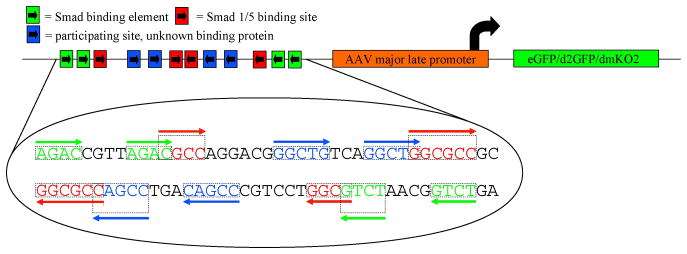
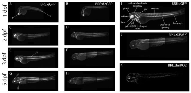
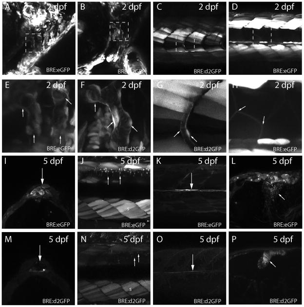
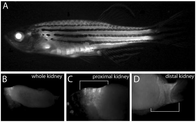
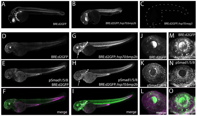
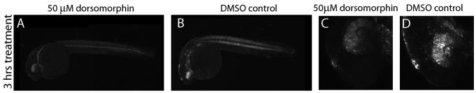
Bone morphogenic protein (BMP) signaling is fundamental to development, injury response, and homeostasis. We have developed transgenic zebrafish that report Smad-mediated BMP signaling in embryos and adults. These lines express either enhanced green fluorescent protein (eGFP), destabilized eGFP, or destabilized Kusabira Orange 2 (KO2) under the well-characterized BMP Response Element (BRE). These fluorescent proteins were found to be expressed dynamically in regions of known BMP signaling including the developing tail bud, hematopoietic lineage, dorsal eye, brain structures, heart, jaw, fins, and somites, as well as other tissues. Responsiveness to changes in BMP signaling was confirmed by observing fluorescence after activation in an hsp70:bmp2b transgenic background or by inhibition in an hsp70:nog3 background. We further demonstrated faithful reportage by the BRE transgenic lines following chemical repression of BMP signaling using an inhibitor of BMP receptor activity, dorsomorphin. Overall, these lines will serve as valuable tools to explore the mechanisms and regulation of BMP signal during embryogenesis, in tissue maintenance, and during disease.


Figures

Figure 1
Figure 1
Schematic of the BRE-reporter construct. Multiple Smad binding elements are arranged in tandem in forward and reverse orientations and placed upstream of the AAV minimal major late promoter (Korchynskyi and ten Dijke, 2002) and used to drive expression of eGFP, destabilized eGFP (d2GFP), or destabilized monomeric Kusabira Orange 2 (dmKO2).
Figure 2
Figure 2
Expression of BRE:d2GFP protein and mRNA, and phospho-Smad1/5/8 immunoreactivty in 13 - 17-somite embryos. Whole embryos were imaged for both d2GFP protein (A, C, E) and mRNA (B, D, F) (green) and phospho-Smad1/5/8 immunoreactivity (magenta) (A’ – F’). Note the enriched and overlapping d2GFP and phospho-Smad1/5/8 immunoreactivity in the tail and lower trunk regions. Higher magnification images of the tailbud region showed co-localization of d2GFP protein and mRNA and phospho-Smad1/5/8 immunoreactivity in some cells (arrowheads), although others expressed only d2GFP protein or mRNA, or phospho-Smad1/5/8. Expression of transcripts for known BMP-regulated targets id1, hey1 and msxE (H, I, J) were found in regions associated with the BRE promoter (G).
Figure 3
Figure 3
Expression of eGFP, d2GFP or KO2-PEST driven by the BRE promoter lines during larval development. BRE:eGFP was expressed in the dorsal developing eye (e), somites (s), tail (t), heart (h) and pineal (p) during the first 5 days post fertilization (A, C, E, G). Similarly, d2GFP was found in the same areas, but at lower levels (B, D, F, H). Annotated expression of enlarged view of 3 dpf BRE:eGFP transgenic zebrafish larvae (I), BRE:d2GFP transgenic zebrafish larvae (J), and BRE:KO2-PEST transgenic zebrafish larvae (K). Montaging artifacts are visible in the trunk region at the border of adjacent image frames.
Figure 4
Figure 4
Diverse tissue expression of eGFP or d2GFP in BRE transgenic lines. (A) Expression of eGFP in cranial vasculature of 2 dpf BRE:eGFP larvae. (B) Similar expression of d2GFP in 2 dpf cranial vasculature of BRE:d2GFP larvae. (C) Expression of d2GFP in intersomitic vasculature of 2 dpf BRE:d2GFP larvae. (D) Expression of eGFP in 2 dpf somite muscle cells and underlying notochord cells of BRE:eGFP larvae. (E–H) Higher magnification of boxed regions in A – D. Arrows indicate the vascular structures in E, F, and G; and the somite muscle cells in H. (I) Expression of eGFP in pineal cells of 5 dpf BRE:eGFP larvae (arrow). (J) Expression of eGFP in somite muscle (s) and dorsal spinal neurons (arrows) of 5 dpf BRE:eGFP larvae. (K) Expression of eGFP in medial longitudinal fasciculus axons (arrow) of 5 dpf BRE:eGFP larvae. (L) Expression of eGFP in midbrain-hindbrain neurons (arrow) of 5 dpf BRE:eGFP larvae. Note the ependymal cells that are also weakly eGFP-positive (asterisks). (M) Expression of d2GFP in pineal cells of 5 dpf BRE:d2GFP larvae (arrow). (N) Expression of d2GFP in somite muscle (s) and dorsal spinal neurons (arrows) of 5 dpf BRE:eGFP larvae. (O) Expression of d2GFP in medial longitudinal fasciculus axons (arrow) of 5 dpf BRE:d2GFP larvae. (P) Expression of d2GFP in hindbrain neural progenitor cells (arrow) of 5 dpf BRE:d2GFP larvae.
Figure 5
Figure 5
BRE:eGFP in adult fish. (A) eGFP expression viewed in whole fish. (B) Dissected kidney showing eGFP expression (C) Higher magnification of proximal and (C) distal regions (white brackets).
Figure 6
Figure 6
Expression of eGFP in regenerating fins of BRE:eGFP adult fish. (A–F) BRE:eGFP expression in adult caudal fins before and during regeneration. Times post-amputation are indicated above each image. Note that BRE:eGFP expression is increased in the regenerating fin tissue following amputation. (G) Magnified view of 7 day post-amputation blastemal cells. (H–N) Corresponding transmitted light images are shown below.
Figure 7
Figure 7
Adult ocular BRE:eGFP expression revealed in cryosections. (A) BRE:eGFP expression was obvious in the lens and dorsal ciliary margin, but not in the central retina (r). (B) Transmitted light image of A. (C) BRE:eGFP expression in the ciliary marginal zone (cmz) and anterior stromal and vascular cells (as, arrows). (D) Transmitted light image of C. (E) BRE:eGFP expression in dorsal retina showing Müller glia (mg) and scleral cells (s). (F) Transmitted light image of E. (G) BRE:eGFP expression in central retina showing choroidal rete vascular cells (cv). Note the lack of glial expression in the central retina. (H) Transmitted light image of G. Scale bars = 200 μm (A, B), 50 μm (C – H).
Figure 8
Figure 8
BRE:eGFP expression in adult dorsal Müller glia. (A) Central transverse cryosection of an adult BRE:eGFP eye. Note that the lens was not imaged as part of the montage. Müller glia (mg); optic nerve head (onh). Region showing eGFP-positive Müller glia is indicated with a white bracket. (B) Higher magnification of BRE:eGFP in dorsal Müller glia. (C) Expression of glutamine synthetase immunoreactivity (anti-GS), a Müller glia marker, in the same section as B. (D) Co-localization of B and C.
Figure 9
Figure 9
BRE:eGFP expression in adult tissues revealed through cryosections. (A) BRE:eGFP expression in the atrium and ventricle of the heart (dotted line) and in the gill filaments. (B) BRE:eGFP expression in the vasculature of the brain. Higher magnification images of the (C) cardiac atrium and (D) gill filaments.
Figure 10
Figure 10
d2GFP levels are altered when BMP activity is manipulated. Overexpression of Bmp2b driven by the inducible hsp70 heat-shock promoter increases d2GFP expression levels (B, E) relative to endogenous BRE:d2GFP in control larvae at 24 hpf when imaged 6 hours after a 30 minute heat shock at 37°C (A, D). Note the loss of the dorsal high-ventral low gradient pattern in the eye which is replaced by a uniformly high level of d2GFP expression. (C) Conversely, inhibition of BMP signaling by hsp70 overexpression of Noggin3 downregulates d2GFP expression. The embryo position is indicated by dashed lines. At 3 dpf, overexpression of Bmp2b increases both d2GFP and pSmad1/5/8 immunoreactivity relative to normal levels, particularly in the jaw (arrow) and pectoral fin (arrowhead) (G, H). In the 3 dpf eye, Bmp2b overexpression increased d2GFP expression and pSmad1/5/8 immunoreactivity (M, N, O).
Figure 11
Figure 11
d2GFP expression driven by the BRE promoter is down-regulated by dorsomorphin. Larvae expressing BRE:d2GFP were treated with DMSO as a control (B, D) or with 50 μM dorsomorphin (A, C) at 24 hpf for 3 hours. d2GFP expression was reduced, particularly in the head and eye following treatment. Magnification: A, B; ×10: C, D; ×40.

References

    1. Banas MC, Parks WT, Hudkins KL, Banas B, Holdren M, Iyoda M, Wietecha TA, Kowalewska J, Liu G, Alpers CE. Localization of TGF-beta signaling intermediates Smad2, 3, 4, and 7 in developing and mature human and mouse kidney. J Histochem Cytochem. 2007;55(3):275–85. - PubMed
    1. Bauer H, Lele Z, Rauch GJ, Geisler R, Hammerschmidt M. The type I serine/threonine kinase receptor Alk8/Lost-a-fin is required for Bmp2b/7 signal transduction during dorsoventral patterning of the zebrafish embryo. Development. 2001;128(6):849–58. - PubMed
    1. Blitz IL, Cho KW. Finding partners: how BMPs select their targets. Dev Dyn. 2009;238(6):1321–31. - PubMed
    1. Connors SA, Trout J, Ekker M, Mullins MC. The role of tolloid/mini fin in dorsoventral pattern formation of the zebrafish embryo. Development. 1999;126(14):3119–30. - PubMed
    1. Dahlqvist C, Blokzijl A, Chapman G, Falk A, Dannaeus K, Ibanez CF, Lendahl U. Functional Notch signaling is required for BMP4-induced inhibition of myogenic differentiation. Development. 2003;130(24):6089–99. - PubMed

Publication types

LinkOut - more resources