. 2012 Jan;33(1):75-88.
doi: 10.1002/hbm.21194. Epub 2011 Feb 17.

Direct optic nerve pulvinar connections defined by diffusion MR tractography in humans: implications for photophobia

Affiliations

Direct optic nerve pulvinar connections defined by diffusion MR tractography in humans: implications for photophobia

Nasim Maleki et al. Hum Brain Mapp. 2012 Jan.

Abstract

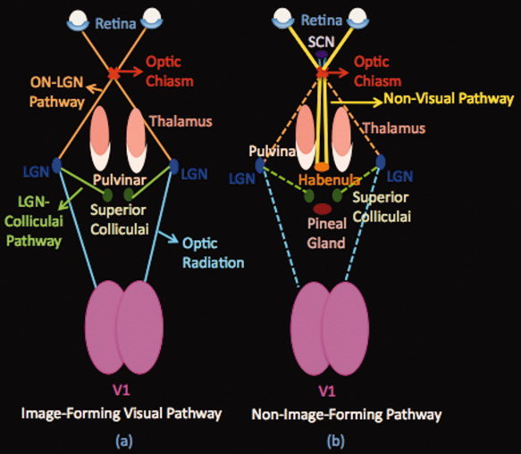
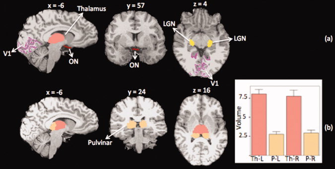
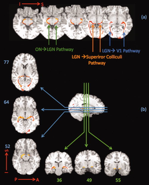
The pathway that underlies exacerbation of migraine headache by light has not been elucidated in the human brain but has recently been reported in a rodent model. We employ diffusion weighted imaging and probabilistic tractography to map connectivity of direct pathways from the optic nerve to the pulvinar implicated with whole-body allodynia during migraine. Nine healthy subjects were recruited to the study and underwent scanning on a 3T magnet. We were able to define well-known image-forming (optic nerve -> lateral geniculate -> visual cortex) as well as a less known nonimage forming visual pathway from the optic chiasm to the pulvinar, and from the pulvinar to several associative cortical brain regions. Such pathway may allow photic signals to converge on a thalamic region we described recently to be selectively activated during migraine headache. Consistent with physiological and anatomical studies in rats, the data provide an anatomical substrate for exacerbation of migraine headache by light in the human.


Figures

Figure 1
Figure 1
Schematic Pathways. (a) Image‐forming visual pathway. This system includes the eye, optic nerve, optic chiasm, lateral geniculate body (LGN), optic radiation, and visual cortex (V1). The pathway starts from the retina of both eyes. The optic nerves of both eyes travel through the optic chiasm to the lateral geniculate nucleus (LGN) of the thalamus (optic tract). From the LGN, a few fibers pass on to the superior colliculus and the rest, optic radiations, continue their way to the occipital lobe terminating in the visual cortex.(b) Nonimage forming Pathway. A specialized pathway involved in nonimage forming functions (entrainment of the biological clock, adaptation of the pupil size to light). This pathway starts from the retina of both eyes as well. The optic nerve fibers project via the retinohypothalamic tract to the suprachiasmatic nucleus (SCN) of the hypothalamus and then project to the habenula and the pineal gland (see text). SCN: Supra‐Chiasmatic Nucleus, LGN: Lateral Geniculate Nucleus.
Figure 2
Figure 2
Masks for tracking visual and nonimage forming pathways. (a) For each subject the following masks were defined to be used in tracking the visual or the nonimage forming pathway: ON: optic nerve mask, V1: Primary visual cortex, LGN: Lateral geniculate nucleus, pulvinar and thalamus. ON mask was used as the seeding mask and the rest of the masks were used as target or exclusion masks. (b) thalamus and pulvinar segmentation results in one subject along with the group average results of the left and right thalamic and pulvinar volumes (graph). The average pulvinar volume was found to be approximately one‐third that of the thalamus. Th: Thalamus, P: Pulvinar, L: Left, and R: Right (example: P‐L: Left Pulvinar)
Figure 3
Figure 3
Image forming visual pathway. The tracking results for the visual system are shown. (a) The pathways connecting the optic nerve to the LGN, optic radiation to the primary visual cortex and those projecting from LGN to the superior colliculi are shown in a single subject. (b) Visual pathway is shown in another subject in axial, sagittal and coronal views. The course of the visual pathway can be followed in these slices from where the optic tract connects the optic nerve to the LGN and then travels to the superior colliculus or the LGN, and next continues to the occipital lobe where it terminates in the visual cortex. The results are consistent with the known visual pathway system.
Figure 4
Figure 4
Nonimage forming visual pathway. (a) A direct pathway that connects the optic nerve to the thalamus. This pathway reaches the posterior thalamic nucleus that includes the pulvinar nucleus. For this analysis, optic chiasm was used as the seeding mask, pulvinar as the target and LGN as an exclusion mask to exclude any visual pathway that goes through the LGN. The pathway shown in this figure is thus nonvisual. Also seen in this figure is the optic chiasm‐hypothalamic connectivity, which presumably represents the suprachiasmatic nucleus projections. (b) (I) Delineation of the non–image forming and visual pathways in an axial slice. The visual and non–image forming pathways are delineated as follows: (i) the LGN‐Colliculi pathway, ON‐Pulvinar pathway and optic radiation are all shown. (ii) Optic‐radiation is excluded by using the V1 mask as an exclusion mask. (iii) The optic radiation and the LGN‐Colliculi pathway are excluded by using the LGN mask and V1 mask as exclusion masks. The green is the LGN‐Colliculi pathway, the blue is the optic radiation and the red is the pathway that connects ON and pulvinar directly. (II) Specificity analysis results. Pathways were tracked from the optic chiasm with pulvinar as the waypoint mask (red‐yellow) or thalamus minus pulvinar as the waypoint mask and pulvinar and LGN as exclusion masks. No pathway was tracked from optic chiasm to any other nuclei of the thalamus. (III) Diffusion tensor color map of an independent DTI study (Wakana et al.,2004 and http://cmrm.med.jhmi.edu/). Arrows refer to fibers passing through the same area as the nonvisual pathway. Red, green, and blue represent fibers running along right‐left, anterior‐posterior, and superior‐inferior axes, respectively.
Figure 5
Figure 5
Pulvinar to brainstem pathway. Pulvinar‐brainstem connecting pathway. This pathway connects the trigeminal nuclei to the pulvinar. This pathway may send nociceptive information originating in trigeminal afferents during migraine via the trigeminal nuclei to the pulvinar.
Figure 6
Figure 6
Cortical‐Pulvinar Pathways. (a) Cortical masks were defined for each subject individually including: Primary motor cortex (M1), olfactory and V1 mask (shown in figure 2a). In Figures 4b–c, group probability maps of reconstructed pathways connecting the pulvinar to (b) M1, and (c) Olfactory cortex are presented. These results are based on tractography in nine subjects. The average tract map is thresholded to show the tracts that are present in at least 50% of the subjects. Yellow represents higher probability of a pathway's presence in more subjects and red is for lower probability (minimum 50%).

References

    1. Amery WK, Waelkens J, Vandenbergh V ( 1988): The sensorium of the migraineur. Ital J Neurol Sci 9: 539–545. - PubMed
    1. Antal A, Lang N, Boros K, Nitsche M, Siebner HR, Paulus W ( 2008): Homeostatic metaplasticity of the motor cortex is altered during headache‐free intervals in migraine with aura. Cereb Cortex 18: 2701–2705. - PubMed
    1. Basser PJ, Pierpaoli C ( 1996): Microstructural and physiological features of tissues elucidated by quantitative‐diffusion‐tensor MRI. J Magn Reson B 111: 209–219. - PubMed
    1. Behrens TE, Berg HJ, Jbabdi S, Rushworth MF, Woolrich MW ( 2007): Probabilistic diffusion tractography with multiple fibre orientations: What can we gain? Neuroimage 34: 144–155. - PMC - PubMed
    1. Behrens TE, Woolrich MW, Jenkinson M, Johansen‐Berg H, Nunes RG, Clare S, Matthews PM, Brady JM, Smith SM ( 2003a): Characterization and propagation of uncertainty in diffusion‐weighted MR imaging. Magn Reson Med 50: 1077–1088. - PubMed

Publication types

LinkOut - more resources