(⁹⁹m)Tc-bisphosphonate-iron oxide nanoparticle conjugates for dual-modality biomedical imaging
- PMID: 21338098
- PMCID: PMC6205601
- DOI: 10.1021/bc100483k
(⁹⁹m)Tc-bisphosphonate-iron oxide nanoparticle conjugates for dual-modality biomedical imaging
Abstract
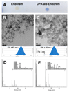
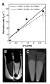
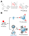
The combination of radionuclide-based imaging modalities such as single photon emission computed tomography (SPECT) and positron emission tomography (PET) with magnetic resonance imaging (MRI) is likely to become the next generation of clinical scanners. Hence, there is a growing interest in the development of SPECT- and PET-MRI agents. To this end, we report a new class of dual-modality imaging agents based on the conjugation of radiolabeled bisphosphonates (BP) directly to the surface of superparamagnetic iron oxide (SPIO) nanoparticles. We demonstrate the high potential of BP-iron oxide conjugation using (⁹⁹m)Tc-dipicolylamine(DPA)-alendronate, a BP-SPECT agent, and Endorem/Feridex, a liver MRI contrast agent based on SPIO. The labeling of SPIOs with (⁹⁹m)Tc-DPA-alendronate can be performed in one step at room temperature if the SPIO is not coated with an organic polymer. Heating is needed if the nanoparticles are coated, as long as the coating is weakly bound as in the case of dextran in Endorem. The size of the radiolabeled Endorem (⁹⁹m)Tc-DPA-ale-Endorem) was characterized by TEM (5 nm, Fe₃O₄ core) and DLS (106 ± 60 nm, Fe₃O₄ core + dextran). EDX, Dittmer-Lester, and radiolabeling studies demonstrate that the BP is bound to the nanoparticles and that it binds to the Fe₃O₄ cores of Endorem, and not its dextran coating. The bimodal imaging capabilities and excellent stability of these nanoparticles were confirmed using MRI and nanoSPECT-CT imaging, showing that (⁹⁹m)Tc and Endorem co-localize in the liver and spleen In Vivo, as expected for particles of the composition and size of (⁹⁹m)Tc-DPA-ale-Endorem. To the best of our knowledge, this is the first example of radiolabeling SPIOs with BP conjugates and the first example of radiolabeling SPIO nanoparticles directly onto the surface of the iron oxide core, and not its coating. This work lays down the basis for a new generation of SPECT/PET-MR imaging agents in which the BP group could be used to attach functionality to provide targeting, stealth/stability, and radionuclides to Fe₃O₄ nanoparticles using very simple methodology readily amenable to GMP.
Figures


References
-
-
For a dedicated recent issue of Chemical Reviews on medical imaging and diagnostics see: Chem Rev. 2010;110(5)
-
-
- Baker M. Whole-animal imaging: The whole picture. Nature. 2010;463:977–80. - PubMed
-
- Pichler BJ, Kolb A, Nagele T, Schlemmer H-P. PET/MRI: Paving the Way for the Next Generation of Clinical Multimodality Imaging Applications. J Nucl Med. 2010;51:333–336. - PubMed
-
- Herzog H, Pietrzyk U, Shah NJ, Ziemons K. The current state, challenges and perspectives of MR-PET. NeuroImage. 2010;49:2072–2082. - PubMed
-
- Wehrl HF, Judenhofer MS, Wiehr S, Pichler BJ. Pre-clinical PET/MR: technological advances and new perspectives in biomedical research. Eur J Nucl Med Mol Imaging. 2009;36:56–68. - PubMed
Publication types
MeSH terms
Substances
Grants and funding
LinkOut - more resources
Full Text Sources
Other Literature Sources
Medical

