. 2011 Feb 25;18(2):252-63.
doi: 10.1016/j.chembiol.2010.12.008.

Heterotaxin: a TGF-β signaling inhibitor identified in a multi-phenotype profiling screen in Xenopus embryos

Affiliations

Heterotaxin: a TGF-β signaling inhibitor identified in a multi-phenotype profiling screen in Xenopus embryos

Michael K Dush et al. Chem Biol. .

Abstract

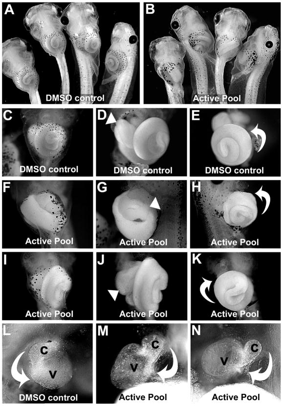
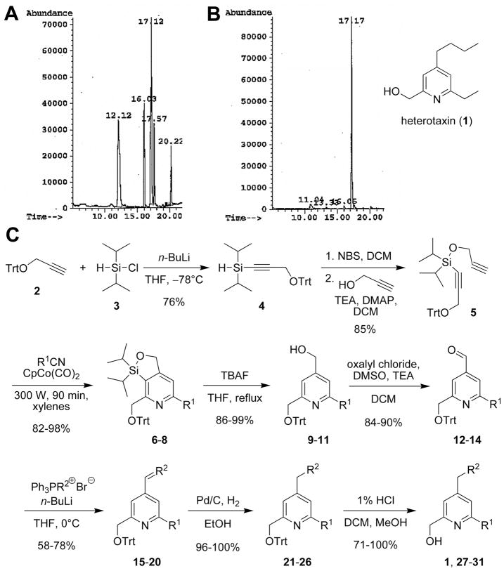
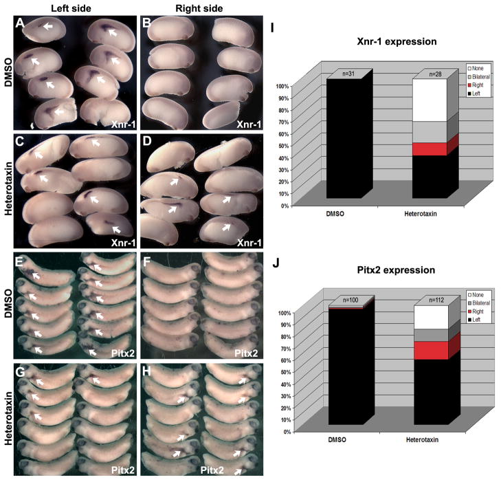
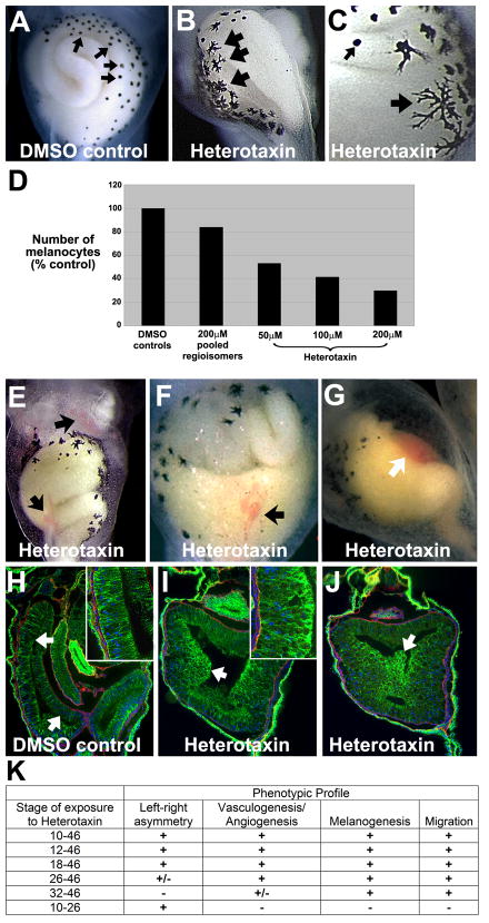
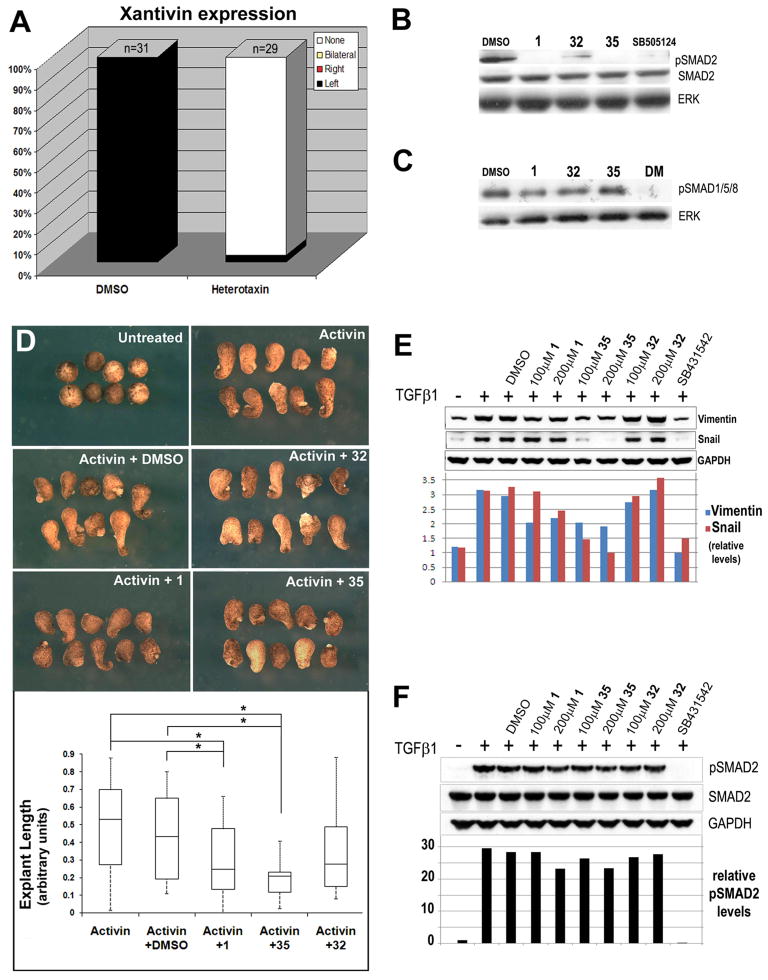
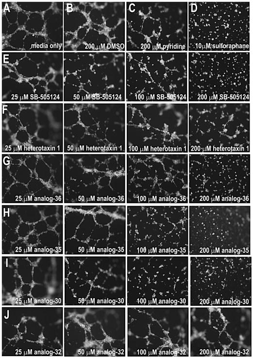
Disruptions of anatomical left-right asymmetry result in life-threatening heterotaxic birth defects in vital organs. We performed a small molecule screen for left-right asymmetry phenotypes in Xenopus embryos and discovered a pyridine analog, heterotaxin, which disrupts both cardiovascular and digestive organ laterality and inhibits TGF-β-dependent left-right asymmetric gene expression. Heterotaxin analogs also perturb vascular development, melanogenesis, cell migration, and adhesion, and indirectly inhibit the phosphorylation of an intracellular mediator of TGF-β signaling. This combined phenotypic profile identifies these compounds as a class of TGF-β signaling inhibitors. Notably, heterotaxin analogs also possess highly desirable antitumor properties, inhibiting epithelial-mesenchymal transition, angiogenesis, and tumor cell proliferation in mammalian systems. Our results suggest that assessing multiple organ, tissue, cellular, and molecular parameters in a whole organism context is a valuable strategy for identifying the mechanism of action of bioactive compounds.


Figures

Figure 1
Figure 1. A mixture of pyridine regioisomers causes heterotaxia in Xenopus
Embryos were treated with A) DMSO or B) 200μM active pool. C) Ventral view of organs in intact DMSO control tadpole. Ventral (D) and left ventral (E) views of tadpole in C with skin removed, showing the normal asymmetry (arrowhead, D) of the foregut loop, and the normal counterclockwise rotation of the intestine (curved arrow, E). F) Tadpole exposed to active pool. Ventral (G) and right ventral (H) view of tadpole in F with skin removed, showing the reversed position (arrowhead, G) of the foregut loop. The intestine is coiling in the normal direction (curved arrow, H), but is located on the opposite side of the animal. Ventral views of tadpole exposed to active pool (I-K), show normal foregut looping (arrowhead, J), but the intestine coiling in the reversed direction (i.e., clockwise; curved arrow, K). L) Ventral view of heart of DMSO control, with outflow tract (conus, c) and ventricle (v) indicated, with the normal direction of cardiac looping (curved arrow). M-N) Two examples of heterotaxin-induced reversed heart looping (curved arrows).
Figure 2
Figure 2. Purification, identification and synthesis of heterotaxin (1)
A) GC trace of the regioisomers in the active pool. B) Active regioisomer (1) after separation (see Supplemental Experimental Procedures), phenotypic assay, and structural assignment. C) Regioselective synthetic route towards gram quantities of heterotaxin (1) and analogs (27–31).
Figure 3
Figure 3. Heterotaxin perturbs left-right asymmetric gene expression patterns
AH) In situ hybridization for nodal (Xnr-1; A-D), and Pitx2 (XPitx2c; E-H) genes was performed on DMSO- (A, B, E, F) and heterotaxin-treated (C, D, G, H) embryos. The expression pattern (arrows) is shown for both the left (A, C, E, G) and right (B, D, F, H) sides of the embryo (A-D, stage 23; E-H, stage 26). The frequency of embryos exhibiting left only (Left, black), right only (Right, red), bilateral (gray) or absent (None, white) expression of nodal (I) or Pitx2 (J) is quantified in the bar graphs.
Figure 4
Figure 4. Heterotaxin perturbs melanogenesis, angiogenesis and cell rearrangements
Compared to the round melanocytes seen in DMSO controls (small arrows, A, C), heterotaxin-treated embryos (two different embryos are shown in B,C) exhibit highly dendritic pigment cells (large arrows, B-C). D) Number of abdominal melanocytes in embryos exposed to either DMSO, the active pool identified in the original pilot screen, or different concentrations of purified heterotaxin. E-G) Heterotaxin induces mild (arrows, E, F) to severe (arrow, G) defects in vasculogenesis (compare the three different examples in E-G to DMSO control in A). H-J) Immunohistochemical staining for E-cadherin, (green), laminin (red) and nuclei (blue; DAPI) on frontal sections (100X) through the gut tube of embryos treated with DMSO (H, control), 100μM heterotaxin (I) or 200μM heterotaxin (J). A single-layer columnar epithelium lines the digestive tract of the DMSO tadpole (arrows, H; inset at 400X), while the cells lining the heterotaxin-treated gut are round, exhibit high levels of E-cadherin (green), and have failed to rearrange into a single layer [arrows, I (inset at 400X), J]. K) Time course studies reveal the sensitivity of each phenotype to heterotaxin exposure. “Left-right asymmetry” = reversal of heart looping, foregut looping, and/or intestinal rotation; Vasculogenesis/Angiogenesis” = hemorrhaging or enlarged blood vessels; “Melanogenesis” = decreased number and increased dendricity of melanocytes; “migration” = perturbed cell rearrangement, indicated by shortening of the primitive gut tube. “+” indicates that at least 75% of embryos exhibited the phenotype in two or more independent trials; “+/−“ indicates that at least 50% of embryos exhibited the phenotype in two or more independent trials. (See Figure S1 for a similar phenotypic profile induced by a known TGF-β inhibitor, SB-505124.)
Figure 5
Figure 5. Synthesis of additional heterotaxin analogs from common precursors
See Table S1 for Structure-Activity Relationship analyses of heterotaxin analogs and Supplemental Experimental Procedures.
Figure 6
Figure 6. Heterotaxin inhibits TGF-β signaling
A) The frequency of embryos exhibiting left only (Left, black), right only (Right, red), bilateral (gray) or absent (None, white) expression of Xantivin is quantified in the bar graphs. (See Figure S2 for images of in situ hybridization). B) Western blotting of embryos exposed from 10 hpf for 24 hrs to DMSO, 200μM heterotaxin (1), 200μM phenotypically inactive analog (32), 80μM active analog (35) and a known nodal signaling inhibitor (SB-505124; 50μM). While unmodified Smad2 is detected under all conditions, phosphorylated Smad2 (pSmad2) is only detected in the presence of DMSO or the inactive heterotaxin analog 32 (albeit at reduced levels). ERK is total protein control. C) Western blotting of embryos exposed to DMSO, 1, 32, 35 and the BMP signaling inhibitor, dorsomorphin (DM; 50μM). BMP-specific Smad1/4/5 levels are unaffected by heterotaxin analogs. D) Activin-induced elongation in Xenopus animal cap explants is inhibited by active heterotaxin analogs 1 and 35 but not DMSO or analog 32. The box plot shows that activin-induced lengthening (results pooled from two identical trials) is inhibited significantly only by compounds 1 and 35, as calculated by one-way ANOVA. *, p<0.05. E) Western blotting of A549 cells exposed to 10ng/ml TGF-β1 and DMSO, 100–200μM 1, 32, 35 or 10μM SB-431542 for 48 hr. TGF-β1-induced upregulation of Vimentin and Snail is inhibited by analogs 1 and 35 but not DMSO or analog 32. GAPDH is total protein control. F) Western blotting of A549 cells exposed to 10ng/ml TGF-β1 and DMSO, 100–200μM 1, 32, 35 or 10μM SB-431542 for 1 hr. TGF-β1-induced phosphorylation of Smad2 is unaffected by heterotaxin analogs. GAPDH controls for total protein. (See Figure S2 for effect of 1 on non-Smad-dependent TGF-β signaling via PI3K/Akt.)
Figure 7
Figure 7. Heterotaxin inhibits angiogenesis in human cells
Human umbilical vein endothelial cells (HUVECs) form tubes when cultured in the presence of media alone (A), DMSO (B), unsubstituted pyridine (C), or the inactive heterotaxin analog 32 (J). In contrast, HUVECs cultured in the presence of a known anti-angiogenic agent (D, sulforophane; Bertl et al., 2006) are unable to form tubes. Although the original heterotaxin molecule 1 (F) exhibits only very mild anti-angiogenic effects in this assay (at 6 hr), the other active heterotaxin analogs 36 (G), 35 (H) and 30 (I) have obvious concentration-dependent anti-angiogenic properties comparable to a known TGF-β signaling inhibitor (E, SB-505124). Cells were stained with Calcein AM to confirm viability. (See Figure S3 for effect of analog 30 on tumor cell growth.)

References

    1. Adams DS, Robinson KR, Fukumoto T, Yuan S, Albertson RC, Yelick P, Kuo L, McSweeney M, Levin M. Early, H+-V-ATPase-dependent proton flux is necessary for consistent left-right patterning of non-mammalian vertebrates. Development. 2006;133:1657–1671. - PMC - PubMed
    1. Bakin AV, Tomlinson AK, Bhowmick NA, Moses HL, Arteaga CL. Phosphatidylinositol 3-kinase function is required for transforming growth factor beta-mediated epithelial to mesenchymal transition and cell migration. J Biol Chem. 2000;275:36803–36810. - PubMed
    1. Bertl E, Bartsch H, Gerhauser C. Inhibition of angiogenesis and endothelial cell functions are novel sulforaphane-mediated mechanisms in chemoprevention. Mol Cancer Ther. 2006;5:575–585. - PubMed
    1. Bertolino P, Deckers M, Lebrin F, ten Dijke P. Transforming growth factor-beta signal transduction in angiogenesis and vascular disorders. Chest. 2005;128:585S–590S. - PubMed
    1. Bols M, Skrydstrup T. Silicon-Tethered Reactions. Chemical Reviews. 1995;95:1253–1277.

Publication types

MeSH terms

LinkOut - more resources