Review
. 2011 Feb;59(2):129-38.
doi: 10.1369/0022155410394857.

Light sheet fluorescence microscopy: a review

Affiliations
Review

Light sheet fluorescence microscopy: a review

Peter A Santi. J Histochem Cytochem. 2011 Feb.

Abstract

Light sheet fluorescence microscopy (LSFM) functions as a non-destructive microtome and microscope that uses a plane of light to optically section and view tissues with subcellular resolution. This method is well suited for imaging deep within transparent tissues or within whole organisms, and because tissues are exposed to only a thin plane of light, specimen photobleaching and phototoxicity are minimized compared to wide-field fluorescence, confocal, or multiphoton microscopy. LSFMs produce well-registered serial sections that are suitable for three-dimensional reconstruction of tissue structures. Because of a lack of a commercial LSFM microscope, numerous versions of light sheet microscopes have been constructed by different investigators. This review describes development of the technology, reviews existing devices, provides details of one LSFM device, and shows examples of images and three-dimensional reconstructions of tissues that were produced by LSFM.


Conflict of interest statement

The author(s) declared no potential conflicts of interest with respect to the authorship and/or publication of this article.

Figures
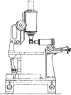
Figure 1.
Figure 1.
This is Figure 3 from Siedentopf and Zsigmondy’s (1903) article and shows part of their light sheet microscope with an upright microscope containing a specimen holder that appears to be mounted to its objective lens and orthogonal illumination at 90° from what appears to be an illuminating objective. The slit aperture and collection of sunlight is shown in another figure (not shown). Magnifications were not provided.
Figure 2.
Figure 2.
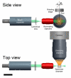
A diagram showing a side and top view of the basic components of a light sheet fluorescence microscope (LSFM). The light sheet is formed by a laser (solid state or gas) and is collimated and expanded with a beam expander. A cylindrical lens forms the light sheet (green beam), and it is projected through an illuminating objective. The focal point or the thinnest portion of the light sheet is positioned usually within the middle of the specimen chamber. The specimen chamber is made of optically clear glass walls and has an open top for specimen insertion. The chamber is filled with either a warmed physiological solution for live-cell imaging or clearing fluid for fixed and cleared tissue. The specimen (white ellipsoid) is attached to a rod and is intersected by the light sheet and a fluorescent plane (i.e., optical section) within the tissue (labeled emission [orange cone]), which is collected by a microscope that is usually mounted in a horizontal position. The specimen rod is attached to rotating and translating stages (not shown) for micropositioning. For a small specimen or a relatively thick light sheet, the fluorescent plane within the tissue is collected by a digital camera as a real-time two-dimensional optical section. However, for specimens larger than the distance of the confocal parameter of the light sheet, the specimen is scanned in the x-axis to produce a well-focused composite image across the width of the specimen. By moving the specimen in the z-axis and collecting another image, a stack of well-aligned, serial optical sections (i.e., a z-stack) through the tissue is obtained. Bar = 5 cm.
Figure 3.
Figure 3.
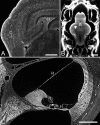
Thin-sheet laser imaging microscopy optical sections from a rat brain, zebrafish head, and mouse cochlea. (A) An optical section from a z-stack from a paraformaldehyde-fixed, unstained rat pup brain (5 weeks old). Neurons are seen as dark structures in the cortex (C) and hippocampus (H) against a lighter background. The image was taken with the line scan camera and adjusted for brightness, contrast, unsharp masking, contrast-limited adaptive histogram equalization, and fast Fourier transform for line shadow reduction. Bar = 500 µm. (B) Rhodamine-stained zebrafish head showing brightly stained bones of the skull (S) and inner ear (I) and lighter staining of the brain (B) with nerve fiber bundles (arrowhead) shown as lightly stained structures. The image was taken with the Retiga CCD camera and adjusted for brightness, contrast, unsharp masking, and fast Fourier transform for line shadow reduction. Bar = 500 µm. (C) Scala media cross section from a mouse cochlea stained with rhodamine showing hair cells (H) of the organ of Corti, tectorial membrane (T), stria vascularis (S), spiral ganglion neurons (SG), and Reissner’s membrane (R). The image was taken with the line scan camera and adjusted for brightness, contrast, and unsharp masking. Bar = 100 µm.
Figure 4.
Figure 4.
Thin-sheet laser imaging microscopy image of the mouse organ of Corti showing outer hair cells labeled with a prestin antibody. (A) The secondary antibody was coupled to the Alexa Fluor 532 fluorophore and showed the presence of the primary antiprestin antibodies within the lateral wall of the outer hair cells where they serve as motor proteins for the outer but not the inner hair cells. (B) An Amira three-dimensional isosurface reconstruction of these prestin-labeled outer hair cells showing the distribution of this protein in the middle but not apical and basal portion of the outer hair cells. The image was taken with the line scan camera and adjusted for brightness and contrast, as well as masked to show only the organ of Corti, and the background was subtracted to remove haze. Bar = 500 µm.

References

    1. Becker K, Jährling N, Kramer ER, Schnorrer F, Dodt HU. 2008. Ultramicroscopy: 3D reconstruction of large microscopical specimens. J Biophotonics. 1:36-42 - PubMed
    1. Buytaert JA, Dirckx JJ. 2007. Design and quantitative resolution measurements of an optical virtual sectioning three-dimensional imaging technique for biomedical specimens, featuring two-micrometer slicing resolution. J Biomed Opt. 12:014039. - PubMed
    1. Denk W, Strickler J, Webb W. 1990. Two-photon laser scanning fluorescence microscopy. Science. 248:73-76 - PubMed
    1. Dodt HU, Leischner U, Schierloh A, Jährling N, Mauch CP, Deininger K, Deussing JM, Eder M, Zieglgsnsberger W, Becker K. 2007. Ultramicroscopy: three-dimensional visualization of neuronal networks in the whole mouse brain. Nat Methods. 4:331-336 - PubMed
    1. Dunsby C. 2008. Optically sectioned imaging by oblique plane microscopy. Opt Express. 16:20306-20316 - PubMed

Publication types