. 2011 Apr 15;286(15):13282-91.
doi: 10.1074/jbc.M110.216226. Epub 2011 Feb 21.

In TNF-stimulated cells, RIPK1 promotes cell survival by stabilizing TRAF2 and cIAP1, which limits induction of non-canonical NF-kappaB and activation of caspase-8

Affiliations

In TNF-stimulated cells, RIPK1 promotes cell survival by stabilizing TRAF2 and cIAP1, which limits induction of non-canonical NF-kappaB and activation of caspase-8

Ian E Gentle et al. J Biol Chem. .

Erratum in

Abstract

RIPK1 is involved in signaling from TNF and TLR family receptors. After receptor ligation, RIPK1 not only modulates activation of both canonical and NIK-dependent NF-κB, but also regulates caspase-8 activation and cell death. Although overexpression of RIPK1 can cause caspase-8-dependent cell death, when RIPK1(-/-) cells are exposed to TNF and low doses of cycloheximide, they die more readily than wild-type cells, indicating RIPK1 has pro-survival as well as pro-apoptotic activities. To determine how RIPK1 promotes cell survival, we compared wild-type and RIPK1(-/-) cells treated with TNF. Although TRAF2 levels remained constant in TNF-treated wild-type cells, TNF stimulation of RIPK1(-/-) cells caused TRAF2 and cIAP1 to be rapidly degraded by the proteasome, which led to an increase in NIK levels. This resulted in processing of p100 NF-κB2 to p52, a decrease in levels of cFLIP(L), and activation of caspase-8, culminating in cell death. Therefore, the pro-survival effect of RIPK1 is mediated by stabilization of TRAF2 and cIAP1.


Figures

FIGURE 1.
FIGURE 1.
TRAF2 and cIAP1 are degraded in RIPK1−/− MEFs in response to TNF. A, wild-type, RIPK1−/−, and TRAF2−/− MEFs were treated with 100 ng/ml Fc-TNF for 1 h and then lysed with DISC buffer. Proteins were analyzed by Western blot for cIAP1, TRAF2, RIPK1, and cFLIP. B, wild-type and RIPK1−/− MEFs were treated with 100 ng/ml Fc-TNF for 0, 15, 30, and 60 min. Cells were then lysed in DISC lysis buffer and split into soluble and insoluble fractions. 25 mg of protein from both fractions was run on SDS-PAGE and analyzed for levels of indicated proteins by Western blot. C, RIPK1−/− MEFs were treated with either 10 μm MG132, 100 mm NH4Cl, or 200 mm chloroquine for 1 h followed by treatment with 100 ng/ml Fc-TNF for 1 h. DISC lysates were then separated into soluble and insoluble and levels of indicated proteins detected by Western blot. D, wild-type and RIPK1−/− MEFs were treated with either 500 nm compound A or untreated for 1 h followed by addition of 100 ng/ml Fc-TNF for 1 h. DISC lysates were made and the soluble and insoluble fraction was analyzed for levels of cIAP1 and TRAF2 by Western blot.
FIGURE 2.
FIGURE 2.
TRAF2 and cIAP1 are degraded in primary tissues treated with TNF. A, E19 embryos were taken and thymus was harvested. Keratinocytes were isolated from day old RIPK−/− 1-day-old pups. Thymi were dispersed into single cell suspensions and half of each was left untreated while the other half was treated with 100 ng/ml Fc-TNF for 1 h. Cells were lysed in DISC lysis buffer and analyzed for TRAF2 and cIAP1 by Western blot. Genotypes were confirmed by Western blot and PCR. B, primary keratinocytes were trated with 100 ng/ml Fc-TNF for 1 h or 500 nm compound A as a positive control for cIAP1 degradation, followed by lysis in DISC buffer and proteins detected by Western blot. C, D645 and HT29 cells were pretreated with 500 nm geldanamycin for 16 h followed by treatment with 100 ng/ml Fc-TNF for 1 h followed by lysis in DISC buffer and analyzed for protein levels by Western blot. D, HT29 cells were infected an lentiviral shRNA against RIPK1 or a control shRNA, and cells were then treated with 100 ng/ml Fc-TNF for 1 h followed by lysis in DISC buffer and analyzed for protein levels by Western blot.
FIGURE 3.
FIGURE 3.
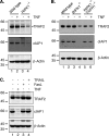
TNFR1 mediates the degradative signal for TRAF2 and cIAP1. A, wild-type and RIPK1−/− MEFs were left untreated or treated with 100 ng/ml soluble mmTNF for 1 h and DISC lysates analyzed for the levels of TRAF2 and cIAP1. B, wild-type, RIPK1−/−, and RIPK1−/− TNFR1−/− double knock-out MEFs were treated with 100 ng/ml Fc-TNF for 1 h and levels of TRAF2 and cIAP1 were assessed. C, wild-type and RIPK1−/− MEFs were treated with either 10 ng/ml of FasL or 1 mg/ml of Iso Leu TRAIL for 1 h, and lysates were analyzed for levels of TRAF2 and cIAP1.
FIGURE 4.
FIGURE 4.
The kinase activity of RIPK1 is not required for protection of TRAF2 and cIAP1. A, wild-type and RIPK1−/− MEFs were pretreated with NEC-1 followed by treatment with 100 ng/ml Fc-TNF for 1 h and lysed in DISC followed by detection of TRAF2 levels by Western blot. B, wild-type and RIPK1−/− MEFs were treated with NEC-1 for 1 h prior to treatment with 100 ng/ml Fc-TNF for 24 h. Cells were isolated and stained with propidium iodide and analyzed for uptake by flow cytometry. Error bars show S.E. of at least three independent experiments. To control for NEC-1 activity, L929 cells.
FIGURE 5.
FIGURE 5.
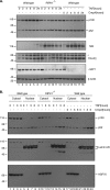
TNF induces non-canonical NF-κB in RIPK1−/− MEFs. A, wild-type and RIPK1−/− MEFs were treated with 100 ng/ml Fc-TNF for the times shown. In parallel, wild-type MEFs were also treated with 500 nm compound A for the same times. Cells were lysed in DISC buffer and analyzed for the levels of NIK, cIAP1, and TRAF2, and processing of p100 to p52. B, wild-type and RIPK1−/− MEFs were treated with 100 ng/ml of Fc-TNF for the indicated times followed by fractionation into cytosolic and nuclear fractions. Fractions were then analyzed by Western blot for the levels of p100/p52 and Lamin A/C and Hsp70 as a loading controls.
FIGURE 6.
FIGURE 6.
Loss of TRAF2 is responsible for the sensitivity of RIPK1−/− MEFs to TNF. Wild-type, RIPK1−/−, and TRAF2−/− MEFs were left untreated or treated with 100 ng/ml Fc-TNF for 24 h. 250 ng/ml CHX was added immediately, 4 h or 8 h after TNF addition, and cells were analyzed for viability by PI uptake using flow cytometry.
FIGURE 7.
FIGURE 7.
RIPK1−/− and TRAF2−/− MEFs transcriptionally up-regulate a pro-survival protein, and show activation of caspase-8. A, wild-type, RIPK1−/−, and TRAF2−/− MEFs were treated with either 100 ng/ml Fc-TNF for 6 h or 100 ng/ml Fc-TNF plus 250 ng/ml of CHX for 6 h. Cells were lysed in DISC buffer and cleavage of caspase-8 and PARP were analyzed by Western blot. B, wild-type, RIPK1−/−, and TRAF2−/− MEFs were treated with either 250 ng/ml of CHX or 25 ng/ml of actinomycin D in combination with 100 ng/ml Fc-TNF for 2 or 4 h. As a control, cells were also treated for 24 h with 100 ng/ml Fc-TNF, 250 ng/ml of CHX, and 25 ng/ml of actinomycin D alone. Cells were harvested and stained with PI followed by analysis by flow cytometry. Error bars show S.E. of at least three independent experiments.
FIGURE 8.
FIGURE 8.
cFLIPL is destabilized in RIPK1−/− and TRAF2−/− MEFs. A, wild-type, RIPK1−/−, and TRAF2−/− MEFs were treated either with MG132 for 1 h prior to treatment with 100 ng/ml Fc-TNF for 1 h, or 100 ng/ml Fc-TNF for 1 h alone. Cells were lysed in DISC buffer and levels of cFLIPL and p43cFLIP were detected by Western blot. B, wild-type, RIPK1−/−, and TRAF2−/− MEFs were treated with either 100 ng/ml Fc-TNF, 250 ng/ml CHX, or a combination of both for the indicated times. Levels of cFLIP and cLFIP-43 were detected by Western blot. C, wild-type, RIPK1−/−, and TRAF2−/− MEFs were infected with 4HT inducible lentiviral constructs coding for cFLIPL or p43cFLIP. Constructs were either induced or not overnight and then treated with 100 ng/ml Fc-TNF for 24 h. Cells were harvested and stained with PI and analyzed for uptake by flow cytometry. Error bars represent S.E. from at least three independent experiments.
FIGURE 9.
FIGURE 9.
Model of pro-death and pro-survival functions of RIPK1. A, wild-type cells. Step 1, TNF binding triggers assembly of complex I by recruitment of TRAFs and cIAPs, which results in p65/RelA NF-κB translocation to the nucleus. Step 2, cFLIPL is up-regulated by NF-κB. Step 3, in the presence of RIPK1, TRAF2, and cIAP1 are stabilized (potentially by direct interaction with RIPK1, but it does not require RIPK1 kinase activity) and TRAF2 mediates stabilization of cFLIPL by an unknown mechanism. Step 4, cFLIPL binds to caspase-8, blocking its activation and preventing apoptosis. B, pro-survival effect of RIPK1. Step 1, TNF binding triggers assembly of complex I by recruitment of TRAFs and cIAPs which results in NF-κB translocation to the nucleus. Step 2, cFLIPL is up-regulated by NF-κB. Step 3, in the absence of RIPK1, cIAP1 and TRAF2 are degraded by a proteasomal/lysosomal mechanism. This also leads to NIK stabilization and p100 processing (not shown), and translocation of p52/RelB dimers to the nucleus. Step 4, loss of TRAF2 leads to destabilization of cFLIPL, the majority of which is degraded by a proteasomal mechanism. A proportion of cFLIPL interacts with caspase-8 and is cleaved to p43cFLIP, thereby blocking full caspase-8 activation. Step 5, partial inhibition of transcription or translation reduces levels of cFLIPL and p43cFLIP allowing caspase-8 activation, resulting in apoptosis.

References

    1. Wong W. W., Gentle I. E., Nachbur U., Anderton H., Vaux D. L., Silke J. (2010) Cell Death Differ. 17, 482–487 - PubMed
    1. Kelliher M. A., Grimm S., Ishida Y., Kuo F., Stanger B. Z., Leder P. (1998) Immunity 8, 297–303 - PubMed
    1. Vince J. E., Pantaki D., Feltham R., Mace P. D., Cordier S. M., Schmukle A. C., Davidson A. J., Callus B. A., Wong W. W., Gentle I. E., Carter H., Lee E. F., Walczak H., Day C. L., Vaux D. L., Silke J. (2009) J. Biol. Chem. 284, 35906–35915 - PMC - PubMed
    1. Mahoney D. J., Cheung H. H., Mrad R. L., Plenchette S., Simard C., Enwere E., Arora V., Mak T. W., Lacasse E. C., Waring J., Korneluk R. G. (2008) Proc. Natl. Acad. Sci. U.S.A. 105, 11778–11783 - PMC - PubMed
    1. Bertrand M. J., Milutinovic S., Dickson K. M., Ho W. C., Boudreault A., Durkin J., Gillard J. W., Jaquith J. B., Morris S. J., Barker P. A. (2008) Mol. Cell 30, 689–700 - PubMed

Publication types

MeSH terms

LinkOut - more resources