. 2011 Feb 21;192(4):647-61.
doi: 10.1083/jcb.201007161.

The frontotemporal dementia mutation R406W blocks tau's interaction with the membrane in an annexin A2-dependent manner

Affiliations

The frontotemporal dementia mutation R406W blocks tau's interaction with the membrane in an annexin A2-dependent manner

Anne Gauthier-Kemper et al. J Cell Biol. .

Abstract

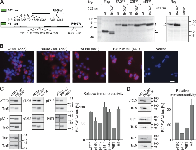
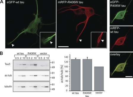
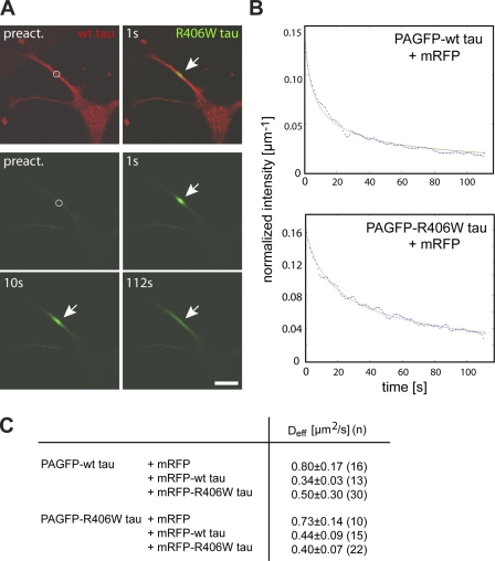
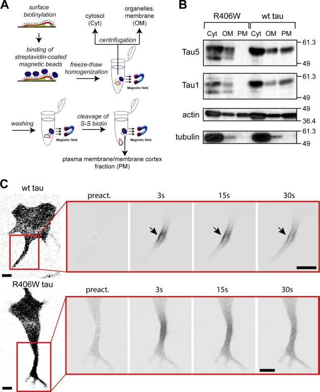
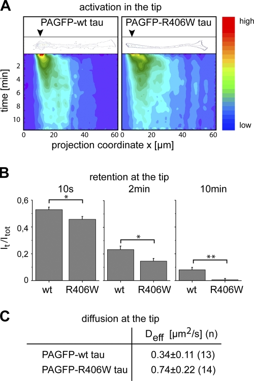
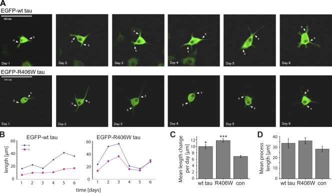
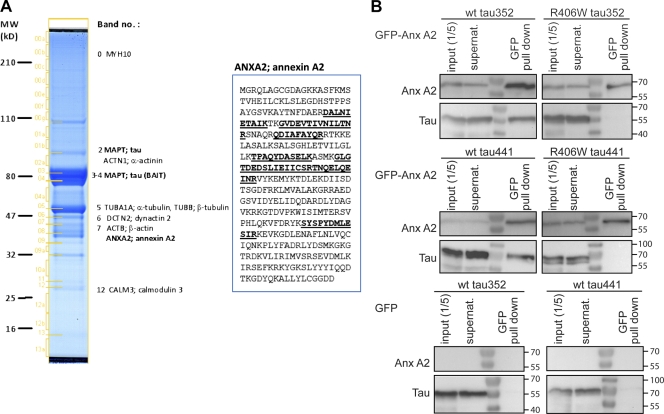
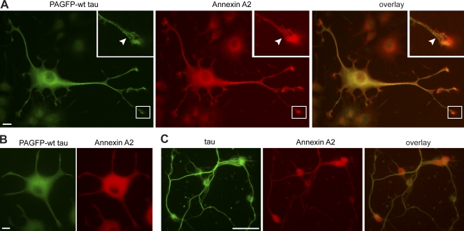
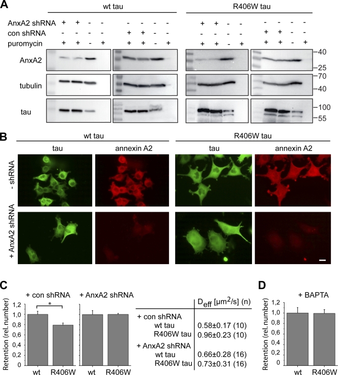
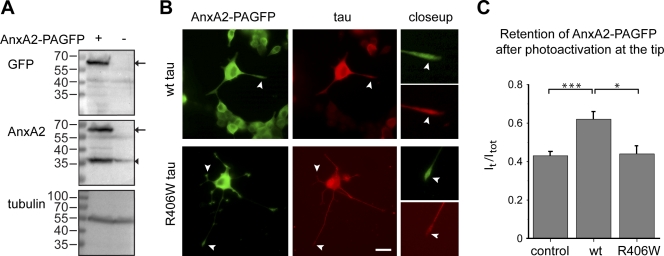
Changes of the microtubule-associated protein tau are central in Alzheimer's disease (AD) and frontotemporal dementia with Parkinsonism linked to chromosome 17 (FTDP-17). However, the functional consequence of the FTDP-17 tau mutation R406W, which causes a tauopathy clinically resembling AD, is not well understood. We find that the R406W mutation does not affect microtubule interaction but abolishes tau's membrane binding. Loss of binding is associated with decreased trapping at the tip of neurites and increased length fluctuations during process growth. Tandem affinity purification tag purification and mass spectrometry identify the calcium-regulated plasma membrane-binding protein annexin A2 (AnxA2) as a potential interaction partner of tau. Consistently, wild-type tau but not R406W tau interacts with AnxA2 in a heterologous yeast expression system. Sequestration of Ca(2+) or knockdown of AnxA2 abolishes the differential trapping of wild-type and R406W tau. We suggest that the pathological effect of the R406W mutation is caused by impaired membrane binding, which involves a functional interaction with AnxA2 as a membrane-cytoskeleton linker.


Figures

Figure 1.
Figure 1.
Expression and phosphorylation of human wt and R406W mutated tau in neural PC12 cells. (A) Schematic representation of the epitope-tagged tau fusion constructs and immunoblot of tau-expressing cells. The tag is indicated as a green flag, the microtubule-binding region (MBR) as black box. Adult-specific exons 2, 3, and 10 are indicated in gray. The R406W FTDP-17 mutation and some disease-relevant phosphorylation sites are indicated (numbering according to the longest central nervous system tau isoform containing 441 aa). The AT270 antibody recognizes phosphorylated T181, PHF1 phosphorylated S396/S404, and Tau1 dephosphorylated S199. Immunoblots of lysates detected with Tau5 antibody show separation of the fusion proteins at the expected size. Note that wt tau separates as a single band (arrowhead), whereas R406W mutant tau exhibits a doublet (arrowhead and arrow) indicative of the presence of a population with decreased phosphorylation. (B) Fluorescence micrographs of PC12 cells stably expressing different tau constructs and a vector control. Proteins were detected using monoclonal anti-FLAG antibody, and nuclei were stained with DAPI. Bar, 10 µm. (C) Immunoblots and phosphorylation profile of tau as detected with a panel of phosphorylation-sensitive antibodies. Lysates were prepared from stably transfected FLAG-tau (352 tau)-expressing cells. Immunoblots are shown for different phosphorylation-sensitive antibodies as indicated (top) and phosphorylation-insensitive Tau5 antibody (bottom). Relative immunoreactivity against individual phosphoepitopes was calculated from the total intensities of all tau-reactive bands per lane divided by Tau5 immunoreactivity. To plot a phosphorylation profile, the respective values for R406W tau were expressed relative to wt tau, which was set as 100%. Mean and range from two independent tau-expressing PC12 cell clones are shown (error bars). 25 µg of protein were loaded per lane. Note the decreased phosphorylation of mutant tau at several sites (T205, T212, and S396/404). The amount of endogenous tau was below the detection limit. Black lines indicate that intervening lines have been spliced out. (D) Immunoblots and relative immunoreactivity of 441 tau, as detected with a selection from the antibodies shown in C. Lysates were prepared from stably transfected Flag-tau (441 tau)-expressing cells. Phosphorylation was decreased at T205, T212, and at the PHF1-epitope, similar to 352 tau. Numbers to the sides of the gel blots indicate molecular mass standards in kilodaltons.
Figure 2.
Figure 2.
Fluorescent-tagged wt and R406W tau interact similarly with microtubules. (A) Fluorescence micrographs (left) and double fluorescence images (right) of detergent-extracted cells expressing the indicated tau constructs. The growth cone region is shown enlarged in the insets. Note the filamentous staining patterns in the growth cone regions (arrowheads) indicative of cytoskeletal association (left). Colocalization of wt tau and R406W tau suggests binding to the same subpopulation of microtubules (right). Bars, 20 µm. (B) Immunodetection of tau, acetylated tubulin (ac-tub), and total α-tubulin of lysates from tau-expressing and vector-control PC12 cell lines. Indicated amounts of protein were loaded per lane. Relative immunoreactivity of the ac-tub signal to total α-tubulin was calculated from the lanes where 10 µg of protein had been separated. Note the increased ratios of acetylated to total tubulin in human tau-expressing cells compared with control lines indicative of microtubule stabilization. No difference was observed between wt and R406W tau–expressing cells. Mean and range from two independent tau-expressing PC12 cell clones are shown (error bars). Experiments were performed with the 352 tau isoform. Numbers to the sides of the gel blots indicate molecular mass standards in kilodaltons.
Figure 3.
Figure 3.
Wt and R406W tau exhibit same effective diffusion in the neuritic shaft. (A) Live cell imaging of a PC12 cell coexpressing mRFP-tagged wt tau and PAGFP-tagged R406W tau after fluorescence photoactivation. The position of photoactivation is indicated by a circle in the preactivation images, and distribution between 1 and 112 s was determined. Fluorescence decay of PAGFP-R406W tau in the activated region as a result of diffusion is marked by the arrows. Bar, 20 µm. (B) Fits of representative decay plots to model diffusion of PAGFP-wt tau and R406W tau. Modeling was performed as described in Materials and methods. (C) Effective diffusion coefficients for PAGFP-tau in different combinations of coexpression. Stably transfected PAGFP-tau–expressing lines were transiently transfected with mRFP or mRFP-tagged constructs as indicated. Coexpression of tau constructs reduces the diffusion similarly for wt and R406W tau. Values are shown as mean ± SEM from fits of n processes. Experiments were performed with the 352 tau isoform.
Figure 4.
Figure 4.
R406W tau is deficient in binding to the neural plasma membrane. (A) Schematic representation of the plasma membrane fractionation assay to analyze tau’s interaction with the neural membrane cortex. (B) Immunoblot showing the distribution of FLAG-tagged tau, actin, and tubulin in the cytosolic (Cyt), organelles/membrane (OM), and plasma membrane/membrane cortex (PM) fractions. Note the complete absence of R406W tau mutant in the PM fraction, whereas a major amount of wt tau is PM-associated. Numbers to the sides of the gel blots indicate molecular mass standards in kilodaltons. (C) Time-lapse microscopic images of processes from PC12 cells expressing PAGFP-tagged tau constructs after photoactivation. A close-up of the processes (red box) is shown and the position of photoactivation is indicated by a circle. Note the enrichment of wt tau close to the plasma membrane (arrows), whereas R406W tau shows a uniform distribution. Experiments were performed with the 352 tau isoform. Bar, 10 µm.
Figure 5.
Figure 5.
R406W tau exhibits reduced trapping in the tip of neurites compared with wt tau. (A) Contour and color-coded plots of 2D intensity functions after photoactivation in the tip of representative processes expressing PAGFP wt and R406W tau. Position of activation is indicated by black arrowheads in the contour plot. Fluorescence intensity is color-coded from blue to red as indicated on the right. Note that the dissipation of fluorescence in the activated region occurs faster with R406W tau compared with wt tau. (B) Quantification of retention after focal activation of wt and R406W tau in the tip of neurites. Immobile fractions at different time points after activation (It/Itot) show decreased retention of R406W compared with wt tau. Values are shown as mean ± SEM (error bars). **, P < 0.01; *, P < 0.05 (n = 12–21). (C) Effective diffusion coefficients of tau after photoactivation at the tip of processes in PC12 cells stably expressing PAGFP wt tau or R406W tau. Values are shown as mean ± SEM with fits from n cells. Note the decreased Deff value corresponding to the increased retention of wt tau compared with R406W tau. Experiments were performed with the 352 tau isoform.
Figure 6.
Figure 6.
The R406W mutation induces higher fluctuations during process growth. (A) Live imaging of PC12 cells stably expressing EGFP-tagged wt tau and R406W tau. Fluorescence micrographs of the same cell at different times after induction of differentiation with NGF are shown. Two representative processes per cell are followed over time and are indicated by arrowheads. Bar, 100 µm. (B) Plot of the lengths of the representative processes indicated in A over time. Note that the processes of the R406W tau–expressing cell showed large fluctuations in lengths over time. (C) Quantification of mean length change per day for tau-expressing cells compared with an EGFP-expressing control (con). Results are shown as mean ± SEM (error bars). ***, P < 0.001; *, P < 0.05 compared with the EGFP-expressing control (n = 52–81 processes). Two independent clonal lines were used per construct. (D) Mean length of processes after 6 d of NGF treatment as determined by population analysis. 23–31 of processes were measured for the respective cell lines. Mean process lengths ± SEM are given (error bars). Note that the overall process length is similar for tau-expressing cells. Experiments were performed with the 352 tau isoform.
Figure 7.
Figure 7.
Identification of annexinA2 as a putative tau interaction partner. (A) TAP of tau protein. Amino-terminally TAP-tagged 441 tau was purified from SKNBE2 cells, separated by SDS-PAGE, and stained with colloidal Coomassie. The overexpressed tau protein is clearly visible at an apparent molecular mass of 70–80 kD. Copurifying proteins identified by LC-MS/MS are labeled. The sequence coverage for the AnxA2 protein sequence was 23% (indicated by marking sequenced peptides underlined and in boldface). (B) Pull-down of GFP-tagged Anx A2 fusion constructs in yeast. Wt tau coprecipitated after pull-down of GFP–Anx A2. No coprecipitation of R406W tau was observed. In control experiments (pull-down of GFP), wt tau did not coprecipitate. Numbers to the sides of the gel blots indicate molecular mass standards in kilodaltons.
Figure 8.
Figure 8.
Distribution of tau and AnxA2 in PC12 cells and primary cortical neurons. (A) Fluorescence micrographs of neuronally differentiated PC12 cells stably expressing PAGFP wt tau. Cells were fixed with paraformaldehyde and stained against GFP and endogenous AnxA2. The growth cone region is shown enlarged in the insets. Note the enrichment of AnxA2 at the tip of processes and colocalization of tau and AnxA2 in filamentous structures of the growth cone (arrowheads). (B and C) Fluorescence micrographs of PC12 cells stably expressing PAGFP wt tau (B) and primary cortical neurons (C) after a combined detergent extraction–fixation protocol to reveal cytoskeletal association. PC12 cells were stained against GFP and endogenous AnxA2, and cortical neurons were stained against endogenous tau and AnxA2. Note that a significant amount of AnxA2 is retained together with tau in the cell body and neurites of PC12 cells, as well as the axons of primary neurons. Experiments were performed with the 352 tau isoform. Bars: (A and B) 10 µm; (C) 50 µm.
Figure 9.
Figure 9.
Annexin A2 is required for trapping of wt tau in the tip of neurites. (A) Immunoblots demonstrating shRNA-mediated knockdown of AnxA2 in PC12 cells. PC12 cells stably expressing PAGFP wt tau or R406W tau were infected with lentiviral particles coding for AnxA2 shRNA (AnxA2 shRNA) or a control shRNA (con shRNA). Infected cells were selected with puromycin as described in Materials and methods. Annexin A2 protein is reduced by ∼80% in AnxA2 shRNA-treated cells. (B) Immunofluorescence images of PC12 cells stably expressing PAGFP wt tau and R406W tau with or without shRNA-mediated annexin knockdown. Staining was against AnxA2 and the GFP tag. After knockdown, AnxA2 is hardly detectable by immunofluorescence. Bar, 10 µm. (C) Retention of wt tau and R406W tau in the tip of processes after treatment with control shRNA and after shRNA-mediated AnxA2 knockdown. Immobile fractions after activation (It/Itot) were determined after 10 s and expressed relative to wt tau. Values are shown as mean ± SEM (error bars); *, P < 0.05 (n = 10–16). Effective diffusion coefficients of the respective experiments are shown in the table. Values are shown as mean ± SEM with fits from n cells. Note that annexin knockdown abolished the difference in retention and Deff values between wt tau and R406W tau. (D) Retention of wt tau and R406W tau in the tip of processes in the presence of the calcium chelator BAPTA/AM (BAPTA). Immobile fractions were determined as described in C. Note that the different retention of wt tau and R406W tau is abolished in the presence of BAPTA. Values are shown as mean ± SEM (error bars; n = 11–16). Experiments were performed with the 352 tau isoform.
Figure 10.
Figure 10.
Tau expression reduces the mobility of AnxA2 in growth cones. (A) Immunoblot showing the expression of PAGFP-tagged AnxA2 at the expected size (arrows). Endogenous annexin is indicated by the arrowhead. Tubulin is stained as loading control. (B) Fluorescence micrographs of PC12 cells stably expressing FLAG-tagged wt tau or R406W tau after infection with a lentivirus encoding AnxA2–PAGFP. Cells were fixed with paraformaldehyde and stained with antibodies against GFP (for AnxA2–PAGFP) and the FLAG-epitope (for tau). Close-ups of the tips of processes are shown on the right. Note that AnxA2–PAGFP showed an enrichment at the tip of neurites similar to endogenous AnxA2. Wt tau colocalized with AnxA2–PAGFP at the tip while R406W tau was absent (arrowheads). Bar, 10 µm. (C) Retention of AnxA2–PAGFP at the tip of neuronally differentiated PC12 cells stably expressing FLAG-tagged wt tau, R406W tau, or control cells. Note the increased retention of AnxA2–PAGFP* in wt tau, but not in R406W tau–expressing cells or control cells. Values are shown as mean ± SEM (error bars). ***, P < 0.001; *, P < 0.05 (n = 6–27). Experiments were performed with the 352 tau isoform.

References

    1. Aoyagi H., Hasegawa M., Tamaoka A. 2007. Fibrillogenic nuclei composed of P301L mutant tau induce elongation of P301L tau but not wild-type tau. J. Biol. Chem. 282:20309–20318 10.1074/jbc.M611876200 - DOI - PubMed
    1. Biernat J., Gustke N., Drewes G., Mandelkow E.M., Mandelkow E. 1993. Phosphorylation of Ser262 strongly reduces binding of tau to microtubules: distinction between PHF-like immunoreactivity and microtubule binding. Neuron. 11:153–163 10.1016/0896-6273(93)90279-Z - DOI - PubMed
    1. Brajenovic M., Joberty G., Küster B., Bouwmeester T., Drewes G. 2004. Comprehensive proteomic analysis of human Par protein complexes reveals an interconnected protein network. J. Biol. Chem. 279:12804–12811 10.1074/jbc.M312171200 - DOI - PubMed
    1. Brandt R., Léger J., Lee G. 1995. Interaction of tau with the neural plasma membrane mediated by tau’s amino-terminal projection domain. J. Cell Biol. 131:1327–1340 10.1083/jcb.131.5.1327 - DOI - PMC - PubMed
    1. Bunker J.M., Kamath K., Wilson L., Jordan M.A., Feinstein S.C. 2006. FTDP-17 mutations compromise the ability of tau to regulate microtubule dynamics in cells. J. Biol. Chem. 281:11856–11863 10.1074/jbc.M509420200 - DOI - PubMed

Publication types