Comparative Study
. 2011 Mar 8;123(9):979-88.
doi: 10.1161/CIRCULATIONAHA.110.006437. Epub 2011 Feb 21.

Disrupted junctional membrane complexes and hyperactive ryanodine receptors after acute junctophilin knockdown in mice

Affiliations
Comparative Study

Disrupted junctional membrane complexes and hyperactive ryanodine receptors after acute junctophilin knockdown in mice

Ralph J van Oort et al. Circulation. .

Abstract

Background: Excitation-contraction coupling in striated muscle requires proper communication of plasmalemmal voltage-activated Ca2+ channels and Ca2+ release channels on sarcoplasmic reticulum within junctional membrane complexes. Although previous studies revealed a loss of junctional membrane complexes and embryonic lethality in germ-line junctophilin-2 (JPH2) knockout mice, it has remained unclear whether JPH2 plays an essential role in junctional membrane complex formation and the Ca(2+)-induced Ca(2+) release process in the heart. Our recent work demonstrated loss-of-function mutations in JPH2 in patients with hypertrophic cardiomyopathy.

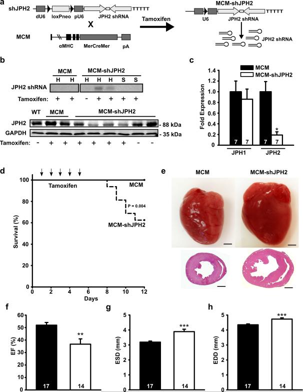
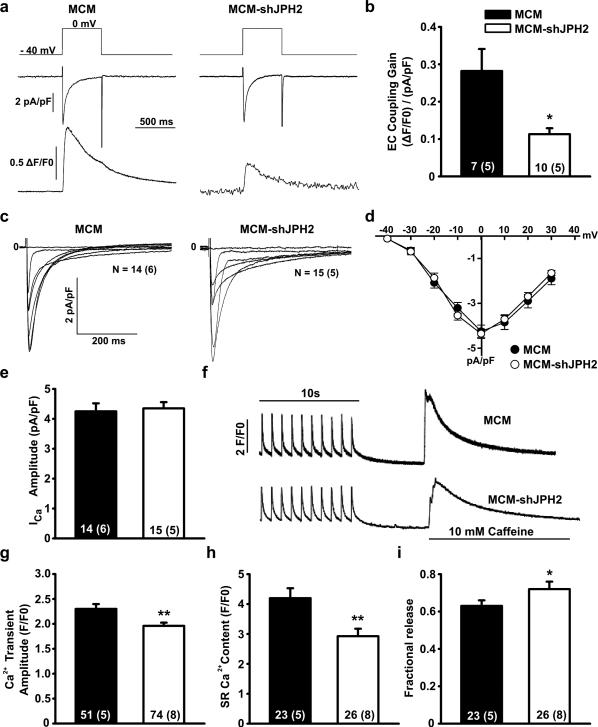
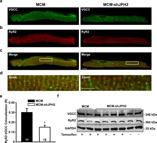
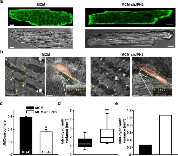
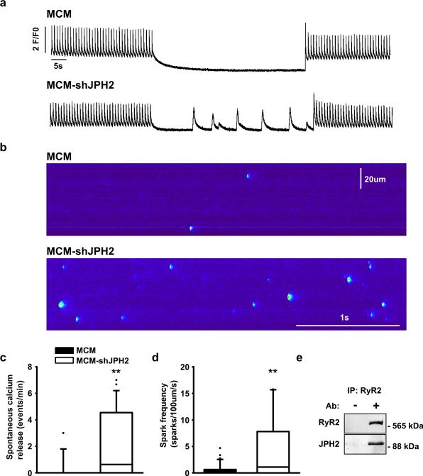
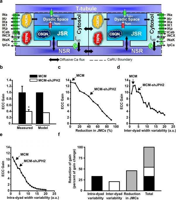
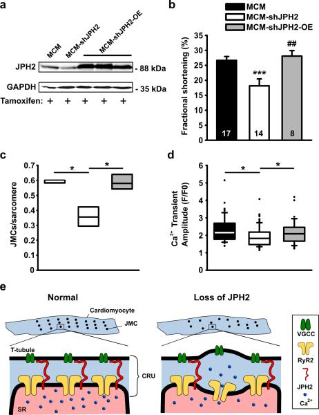
Methods and results: To elucidate the role of JPH2 in the heart, we developed a novel approach to conditionally reduce JPH2 protein levels using RNA interference. Cardiac-specific JPH2 knockdown resulted in impaired cardiac contractility, which caused heart failure and increased mortality. JPH2 deficiency resulted in loss of excitation-contraction coupling gain, precipitated by a reduction in the number of junctional membrane complexes and increased variability in the plasmalemma-sarcoplasmic reticulum distance.

Conclusions: Loss of JPH2 had profound effects on Ca2+ release channel inactivation, suggesting a novel functional role for JPH2 in regulating intracellular Ca2+ release channels in cardiac myocytes. Thus, our novel approach of cardiac-specific short hairpin RNA-mediated knockdown of junctophilin-2 has uncovered a critical role for junctophilin in intracellular Ca2+ release in the heart.


Figures

Figure 1
Figure 1. Inducible cardiac-specific JPH2 knockdown causes mortality and acute heart failure
a. Schematic representation of targeting vector to overexpress JPH2-targeting shRNA sequence (shJPH2) downstream of a U6 promoter, inactivated by insertion of a loxP-flanked neomycin (neo) cassette dividing the distal (dU6) and proximal (pU6) parts of the promoter. Tamoxifen administration to double transgenic offspring of αMHC-MerCreMer (MCM) and shJPH2 mice leads to cardiac-specific Cre-mediated excision of the neo cassette, resulting in expression of JPH2 shRNA. b. Northern blot analysis (top) of JPH2 shRNA in hearts (H) and skeletal muscle (S) and Western blot analysis (bottom) for JPH2 protein levels in heart lysates from tamoxifen-treated MCM-shJPH2 mice. c. Quantitative PCR analysis of JPH1 and JPH2 mRNA levels in cardiac tissue from MCM and MCM-shJPH2 mice. d. Kaplan-Meier curve revealing increased mortality in MCM-shJPH2 compared to MCM mice. Arrows indicate single tamoxifen injections at days 1 to 5. e. Representative whole mounts (top) and H&E-stained transverse sections (bottom) of MCM and MCM-shJPH2 mouse hearts 1 week after completion of tamoxifen administration. Scale bar = 1 mm. f, g and h. Echocardiographic measurement of ejection fraction (EF) (f), end-systolic diameter (ESD) (g), and end-diastolic diameter (EDD) (h) 1 week after tamoxifen. Data are represented as mean ± SEM; * P<0.05, ** P<0.01, *** P<0.001 versus MCM control. Number of mice indicated inside bars.
Figure 2
Figure 2. JPH2 knockdown reduces excitation-contraction coupling by suppression of Ca2+-induced Ca2+ release
a. Representative simultaneous tracings evoked by a 40 mV depolarization step (upper panel) of Ca2+ current through the VGCC (middle panel) and whole cell Ca2+ transient (lower panel). b. Bar graph of excitation-contraction coupling gain. c. Representative whole-cell patch clamp current tracings of amplitude and inactivation kinetics of VGCC. d. Current-voltage curves showing activation kinetics of VGCC. e. Bar graph showing peak amplitude of Ca2+ current through VGCC at 0 mV. f. Representative tracings of Ca2+ transient amplitudes when paced at 1 Hz and total SR Ca2+ content as measured by the amplitude of caffeine-induced Ca2+ transient. g. Bar graph demonstrating decreased average Ca2+ transients in MCM-shJPH2 mice. h. Bar graph showing decreased average SR Ca2+ load in MCM-shJPH2 mice. i. Fractional release (SR Ca2+ transient normalized for SR Ca2+ load) was increased in MCM-shJPH2 mice. Data are represented as mean ± SEM; numbers indicate total number of cells, with number of mice between brackets; * P<0.05, ** P<0.01 versus MCM control.
Figure 3
Figure 3. Impaired co-localization of VGCC and RyR2 in cardiomyocytes following JPH2 knockdown
a-c. Representative examples of immunofluorescent labeling of ventricular myocytes isolated from tamoxifen-treated MCM or MCM-shJPH2 mice for VGCC (a), RyR2 (b), and both channels (c). Scale bar = 10 μm. d. Close-ups from images given in panel c demonstrate decreased overlap between VGCC and RyR2 channels in MCM-shJPH2 myocytes. Scale bar = 5 μm. e. Bar graph showing quantification of VGCC-RyR2 co-localization (R), which was significantly reduced following JPH2 knockdown. f. Western blots showing unaltered expression of VGCC and RyR2 following JPH2 knockdown in MCM-shJPH2 mice. Averaged data are represented as mean ± SEM; numbers indicate total number of cells; * P<0.05 versus MCM control.
Figure 4
Figure 4. JPH2 knockdown causes fewer but more variable junctional membrane complexes
a. Isolated ventricular myocytes stained with di-8-ANEPPS to visualize T-tubule organization and shown in bright filed. Scale bar = 10 μm b. Low magnification electron micrographs of JMCs (yellow arrow heads) in myocytes from MCM-shJPH2 and MCM control mice. Scale bar, 1 μm. High magnification micrographs demonstrate altered morphology of remaining JMCs after JPH2 knockdown (orange pseudocolor indicates SR, and the T-tubule is colored in green). The insets demonstrate more variable distance between SR and T-tubule membranes in MCM-shJPH2 cardiomyocytes (yellow lines). Scale bar, 100 nm. c. Quantification of the number of JMCs per sarcomere. d. Bar graph showing that the average dyad width variance per JMC was larger in myocytes from MCM-shJPH2 mice. e. Bar graph showing overall variance among all measurements of average JMC distance in MCM-shJPH2 versus MCM controls. Averaged data are represented as mean ± SEM; numbers indicate total number of cells, with number of mice between brackets; * P<0.05, ** P<0.01 versus MCM control.
Figure 5
Figure 5. JPH2 knockdown causes defective RyR2 gating resulting in spontaneous SR Ca2+ release
a. Representative Ca2+ transient recordings demonstrating increased incidence of spontaneous SR Ca2+ release events in ventricular myocytes from MCM-shJPH2 mice. b. Confocal microscopy line scans revealing increased number of Ca2+ sparks in MCM-shJPH2 mice. c, d. Box plots showing quantification of the number of spontaneous Ca2+ release events (c, n=26 cells from 4 mice MCM and 13 cells from 5 mice MCM-shJPH) and Ca2+ sparks (d, n=37 cells from 4 mice MCM and 28 cells from 5 mice MCM-shJPH). e. Co-immunoprecipitation of heart lysates from a wild-type mouse in the presence or absence of anti-RyR2 antibody. Western blots showing that immunoprecitation using anti-RyR2 antibody pulled down JPH2. ** P<0.01 versus MCM control.
Figure 6
Figure 6. Computational analysis reveals effect of JMC alterations on EC coupling gain
a. Schematic representation of the novel computational model of discrete Ca2+ release in a cardiomyocyte. The different Ca2+ sources and sinks, as well as the various ion channels and Ca2+ binding proteins incorporated in the model are shown. Excitation-contraction (EC) coupling gain is defined as the amount of Ca2+ released from the sarcoplasmic reticulum (SR) per unit of Ca2+ entering the cell from the extracellular space (T-tubule). b. Comparison of EC coupling gain in myocytes from MCM and MCM-shJPH2 mice, as measured using Ca2+ imaging (see Figure 2B) and predicted by the computational model. c-e. Simulations reveal the effect of a reduced number of junctional membrane complexes (JMCs) (c), an increase in inter-dyad width variability (d), and an increase in intra-dyad width variability (e) on EC coupling gain. Arrows indicate model outcomes for MCM and MCM-shJPH2 mice. f. Bar chart showing the individual and cumulative contributions of each parameter on EC coupling gain.
Figure 7
Figure 7. JPH2 knockdown effects can be rescued by transgenic overexpression of JPH2
a. Western blot demonstrating that crossing MCM-shJPH2 mice with transgenic mice overexpressing JPH2 in the heart (OE) restores JPH2 protein to supranormal levels in tamoxifen-treated mice. b. Fractional shortening measured using echocardiography. Data are represented as mean ± SEM; number of mice inside bars. c. Box plot indicating number of JMCs per sarcomere, as measured by electron microscopy (n=400 sarcomeres per group). d. Box plot of Ca2+ transient amplitudes measured in isolated cardiomyocytes. e. Proposed model of the role of junctophilin-2 (JPH2) in cardiac myocytes. In normal ventricular myocytes (left), junctional membrane complexes (JMCs) are distributed evenly across sarcomeres. Each JMC contains calcium release units (CRU) comprised of voltage-gated Ca2+ channels (VCGC) on the sarcolemmal T-tubules, and intracellular Ca2+ release channels/ ryanodine receptors (RyR2) on the sarcoplasmic reticulum (SR). Loss of JPH2 expression leads to reduced numbers of functional JMCs per sarcomere, increased variability of the distance between the T-tubule and SR membranes, and defective RyR2 inactivation. * P<0.05, *** P<0.001 versus MCM control, # P<0.05, ## P<0.01 vs MCM-shJPH2.

References

    1. Bers DM. Cardiac excitation-contraction coupling. Nature. 2002;415:198–205. - PubMed
    1. Franzini-Armstrong C, Protasi F, Ramesh V. Shape, size, and distribution of Ca2+ release units and couplons in skeletal and cardiac muscles. Biophys J. 1999;77:1528–1539. - PMC - PubMed
    1. Song LS, Sobie EA, McCulle S, Lederer WJ, Balke CW, Cheng H. Orphaned ryanodine receptors in the failing heart. Proc Natl Acad Sci U S A. 2006;103:4305–4310. - PMC - PubMed
    1. Takeshima H, Komazaki S, Nishi M, Iino M, Kangawa K. Junctophilins: A novel family of junctional membrane complex proteins. Mol Cell. 2000;6:11–22. - PubMed
    1. Garbino A, van Oort RJ, Dixit SS, Landstrom AP, Ackerman MJ, Wehrens XH. Molecular evolution of the junctophilin gene family. Physiol Genomics. 2009;37:175–186. - PMC - PubMed

Publication types

MeSH terms