. 2011 Feb 1;2(1):1-17.

Acute Seizures in Old Age Leads to a Greater Loss of CA1 Pyramidal Neurons, an Increased Propensity for Developing Chronic TLE and a Severe Cognitive Dysfunction

Affiliations
Free PMC article

Acute Seizures in Old Age Leads to a Greater Loss of CA1 Pyramidal Neurons, an Increased Propensity for Developing Chronic TLE and a Severe Cognitive Dysfunction

Bharathi Hattiangady et al. Aging Dis. .
Free PMC article

Abstract

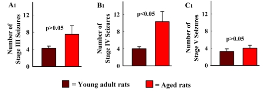
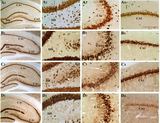
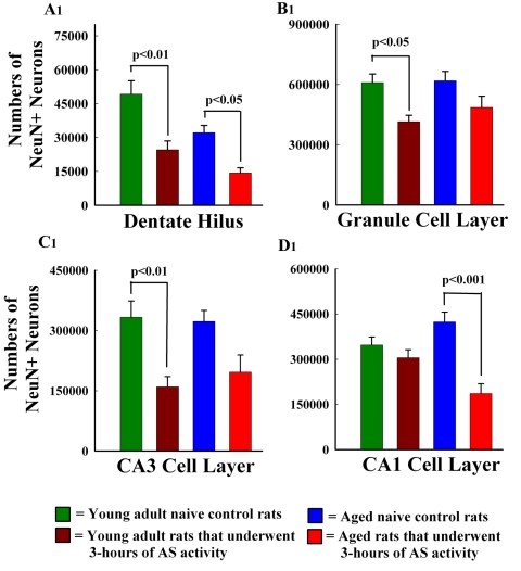
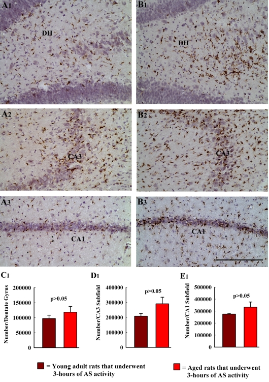
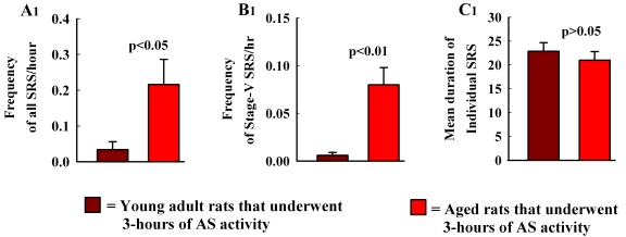
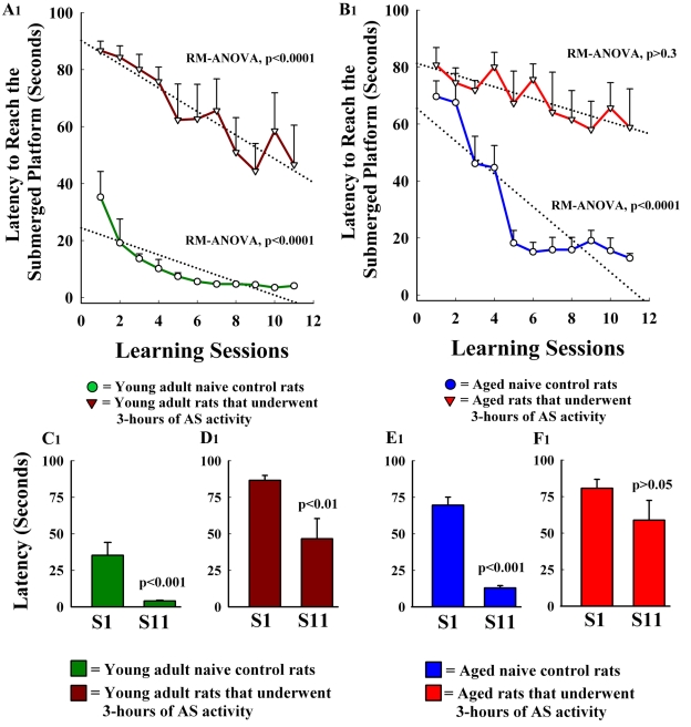
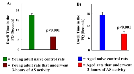
The aged population displays an enhanced risk for developing acute seizure (AS) activity. However, it is unclear whether AS activity in old age would result in a greater magnitude of hippocampal neurodegeneration and inflammation, and an increased predilection for developing chronic temporal lobe epilepsy (TLE) and cognitive dysfunction. Therefore, we addressed these issues in young-adult (5-months old) and aged (22-months old) F344 rats after three-hours of AS activity, induced through graded intraperitoneal injections of kainic acid (KA), and terminated through a diazepam injection. During the three-hours of AS activity, both young adult and aged groups exhibited similar numbers of stage-V motor seizures but the numbers of stage-IV motor seizures were greater in the aged group. In both age groups, three-hour AS activity induced degeneration of 50-55% of neurons in the dentate hilus, 22-32% of neurons in the granule cell layer and 49-52% neurons in the CA3 pyramidal cell layer without showing any interaction between the age and AS activity. However, degeneration of neurons in the CA1 pyramidal cell layer showed a clear interaction between the age and AS activity (12% in the young adult group and 56% in the aged group), suggesting that an advanced age makes the CA1 pyramidal neurons more susceptible to die with AS activity. The extent of inflammation measured through the numbers of activated microglial cells was similar between the two age groups. Interestingly, the predisposition for developing chronic TLE at 2-3 months after AS activity was 60% for young adult rats but 100% for aged rats. Moreover, both frequency & intensity of spontaneous recurrent seizures in the chronic phase after AS activity were 6-12 folds greater in aged rats than in young adult rats. Furthermore, aged rats lost their ability for spatial learning even in a scrupulous eleven-session water maze learning paradigm after AS activity, in divergence from young adult rats which retained the ability for spatial learning but had memory retrieval dysfunction after AS activity. Thus, AS activity in old age results in a greater loss of hippocampal CA1 pyramidal neurons, an increased propensity for developing robust chronic TLE, and a severe cognitive dysfunction.


Figures

Figure 1
Figure 1
Comparison of the numbers of stages III–V seizures during the three hours of acute seizure (AS) activity between the young adult and aged rats. Note that there are no significant differences between the two age groups for the numbers of stage III seizures (A1) or stage V seizures (C1). However, the numbers of stage IV seizures (B1) are greater in aged rats than in young adult rats (p<0.05).
Figure 2
Figure 2
Comparison of the cytoarchitecture of the hippocampus through NeuN immunostaining between a young adult naïve control rat (A1), a young adult rat at 12 days after three hours of acute seizure (AS) activity (B1), an aged naïve control rat (C1), and an aged rat at 12 days after three hours of AS activity (D1). Scale bar, A1–D1 = 500 μm. A2, B2, C2 & D2 illustrate magnified views of the dentate hilus from A1, B1, C1 and D1. Note the loss of dentate hilar neurons in both young adult and aged rats after AS activity (B2, D2) in comparison to age-matched controls (A2, C2). A3, B3, C3 & D3 show enlarged views of the CA3 pyramidal cell layer from the four groups. Note a reduced packing density of neurons in both young adult and aged rats after AS activity (B3, D3), in comparison to respective age-matched controls (A3, C3). A4, B4, C4 & D4 show magnified views of the CA1 pyramidal cell layer from the four groups. In comparison to age-matched controls (A4, C4), thinning of the CA1 pyramidal cell layer is evident in the young adult rat that underwent AS (B4) and a considerable loss of CA1 pyramidal neurons is obvious in the aged rat that underwent AS (D4). Scale bar, A2–D2, A3–D3, A4–D4 = 100 μm.
Figure 3
Figure 3
Comparison of the numbers of NeuN+ neurons between different groups for the dentate hilus (A1), the dentate granule cell layer (B1), the CA3 pyramidal cell layer (C1), and the CA1 pyramidal cell layer (D1). Two-way ANOVA revealed that the loss of neurons after AS between the two age groups was comparable for the dentate hilus (50–55% reduction, p<0.001; A1), the dentate granule cell layer (22–32% reduction, p<0.01; B1) and the CA3 pyramidal cell layer (49–52% reduction, p<0.01; C1). Hence, there was no interaction between the age and AS for the loss of neurons in these regions. In contrast, there was an interaction between age and AS activity for the CA1 pyramidal cell layer (D1) because AS activity induced a differential loss of CA1 pyramidal neurons between young adult rats (12% reduction) and aged rats (56% reduction). Bonferroni post-tests revealed that AS-mediated loss of neurons was significant for the dentate hilus (p<0.01), the granule cell layer (p<0.05) and the CA3 pyramidal cell layer (p<0.01) of young adult rats (A1, B1, C1), and the dentate hilus (p<0.05) and the CA1 pyramidal cell layer (p<0.001) of aged rats (A1, D1).
Figure 4
Figure 4
Comparison of the density and morphology of activated microglial cells (identified through ED-1 immunostaining and hematoxylin counterstaining) in different regions of the hippocampus between the two age groups at 12 days after three hours of AS activity. A1, A2 & A3 respectively illustrate ED-1+ cells (in brown color) in the dentate gyrus (DG), the CA3 subfield and the CA1 subfield of a young adult rat that underwent AS. Whereas, B1, B2 & B3 respectively show ED-1+ elements in the DG, the CA3 subfield and the CA1 subfield of an aged rat that underwent AS. Note that ED-1+ cells in different hippocampal regions of the aged rat (B1–B3) have more complex processes than their counterparts in the young adult rat (A1–A3). Scale bar, A1–B3 = 100 μm. The bar charts in C1, D1 & E1 respectively compare the numbers of ED-1+ activated microglial cells in the DG, the CA3 subfield and the CA1 subfield between the two age groups at 12 days after three hours of AS activity. Note that, the numbers are statistically comparable between the two groups for all regions of the hippocampus.
Figure 5
Figure 5
Comparison of the frequency and intensity of spontaneous recurrent seizures (SRS) between the young adult rats and aged rats in the 2nd and 3rd months after three hours of acute seizure (AS) activity. Figures A1 & B1 compare the frequencies of all SRS (A1) and the stage V seizures (B1; the most severe form of SRS) between the two age groups. Note that in aged rats that underwent three hours of AS activity, the overall frequency of SRS is 6.4 folds greater (p<0.05) and the frequency of stage V seizures is 12.6 folds greater (p<0.01), in comparison to young adult rats that underwent three hours of AS activity. However, the duration of individual SRS between the two age groups was comparable (C1).
Figure 6
Figure 6
Comparison of the spatial learning function in a water maze test between young adult naïve control rats & young adult rats at 3 months after acute seizure (AS) activity (A1), and between aged naïve control rats & aged rats at 3 months after AS activity (B1). Note that, repeated measured ANOVA showed capability for spatial learning in the young adult control group (the green colored learning curve in A1), the aged control group (the blue colored learning curve in B1) and the young adult group that underwent 3-hours of AS activity (the maroon colored learning curve in A1). The Newman-Keuls post-tests revealed a significant decrease in the latency to reach the submerged platform between the first and eleventh sessions in all of these groups (C1, D1 & E1). However, the aged group that underwent 3-hours of AS activity exhibited inability for spatial learning (the red colored learning curve in B1). There was also no significant reduction in the latency to reach the hidden platform between the first and eleventh sessions (F1). Thus, among the four groups of rats, only the aged rats that underwent 3-hours of AS activity exhibited a clear inability for spatial learning.
Figure 7
Figure 7
Comparison of the memory retrieval function between young adult naïve control rats & young adult rats at 3 months after acute seizure (AS) activity (A1), and between aged naïve control rats & aged rats at 3 months after AS activity (B1), as measured through a 45 second probe test conducted at 24 hrs after the last learning session in all four groups. Note that, both young adult control and aged control groups exhibit a clear ability for memory retrieval, which is evidenced by greater dwell times in the platform quadrant (green and blue bars in A1 & B1). In contrast, both young adult and aged rats that underwent AS activity display inability for memory retrieval, which is evidenced by significantly reduced dwell times in the platform quadrant (brown and red bars in A1 & B1).

Similar articles

Cited by

References

    1. Bhatnagar M, Cintra A, Chadi G, et al. Neurochemical changes in the hippocampus of the brown Norway rat during aging. Neurobiol Aging. 1997;18:319–327. - PubMed
    1. Hattiangady B, Rao MS, Shetty GA, Shetty AK. Brain-derived neurotrophic factor, phosphorylated cyclic AMP response element binding protein and neuropeptide Y decline as early as middle age in the dentate gyrus and CA1 and CA3 subfields of the hippocampus. Exp Neurol. 2005;195:353–371. - PubMed
    1. Nicolle MM, Gonzalez J, Sugaya K, Baskerville KA, Bryan D, Lund K, Gallagher M, McKinney M. Signatures of hippocampal oxidative stress in aged spatial learning-impaired rodents. Neuroscience. 2001;107:415–431. - PubMed
    1. Shetty AK, Hattiangady B, Shetty GA. Stem/progenitor cell proliferationfactors FGF-2, IGF-1, and VEGF exhibit early decline during the course of aging in the hippocampus: role of astrocytes. Glia. 2005;51:173–186. - PubMed
    1. Shetty AK, Rao MS, Hattiangady B, Zaman V, Shetty GA. Hippocampal neurotrophin levels after injury: Relationship to the age of the hippocampus at the time of injury. J Neurosci Res. 2004;78:520–532. - PubMed

LinkOut - more resources