. 2010 Dec:25:32-49.
doi: 10.3767/003158510X538371. Epub 2010 Oct 29.

Phytophthora taxa associated with cultivated Agathosma, with emphasis on the P. citricola complex and P. capensis sp. nov

Affiliations

Phytophthora taxa associated with cultivated Agathosma, with emphasis on the P. citricola complex and P. capensis sp. nov

C M Bezuidenhout et al. Persoonia. 2010 Dec.

Abstract

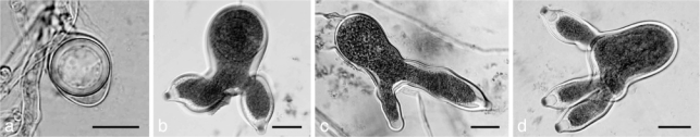
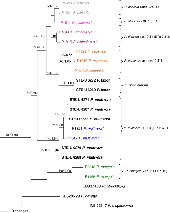
Agathosma species, which are indigenous to South Africa, are also cultivated for commercial use. Recently growers experienced severe plant loss, and symptoms shown by affected plants suggested that a soilborne disease could be the cause of death. A number of Phytophthora taxa were isolated from diseased plants, and this paper reports their identity, mating type, and pathogenicity to young Agathosma plants. Using morphological and sequence data seven Phytophthora taxa were identified: the A1 mating type of P. cinnamomi var. cinnamomi, P. cinnamomi var. parvispora and P. cryptogea, the A2 mating type of P. drechsleri and P. nicotianae, and two homothallic taxa from the P. citricola complex. The identity of isolates in the P. citricola complex was resolved using reference isolates of P. citricola CIT groups 1 to 5 sensu Oudemans et al. (1994) along with multi-locus phylogenies (three nuclear and two mitochondrial regions), isozyme analyses, morphological characteristics and temperature-growth studies. These analyses revealed the isolates from Agathosma to include P. multivora and a putative novel species, P. taxon emzansi. Furthermore, among the P. citricola reference isolates the presence of a new species was revealed, described here as P. capensis. Findings of our study, along with some recent other studies, have contributed to resolving some of the species complexity within the P. citricola complex, resulting in the identification of a number of phylogenetically distinct taxa. The pathogenicity of representative isolates of the taxa from Agathosma was tested on A. betulina seedlings. The putative novel species, P. taxon emzansi, and P. cinnamomi var. parvispora were non-pathogenic, whereas the other species were pathogenic to this host.

Keywords: avocado; buchu; fynbos; glucose-6-phosphate isomerase; isozymes; malate dehydrogenase; pathogenicity; root-rot; taxonomy.


Figures

Fig. 1
Fig. 1
Mean radial growth rates (mm per day) on carrot agar of Phytophthora taxa in the Phytophthora citricola complex. The standard error (SE) is included only for P. taxon emzansi where there were significant differences in growth rate between isolates. All other species in the P. citricola complex had less than 10 % SE at each temperature and therefore error bars are not shown. Green line – P. plurivora (CIT1) (N = 1); orange line – P. citricola clade E sensu Jung & Burgess (2009) (syn CIT2) (N = 2); dark blue line – P. multivora (CIT3) (N = 8); purple line – P. taxon emzansi (CIT4) (N = 2); pink line – P. capensis (CIT4) (N = 3); dashed black line – P. mengei (CIT5) (N = 1), light blue line – P. citricola sensu Oudemans et al. (1994) (CIT1, ET 2 & 3) (N = 2)
Fig. 2
Fig. 2
Morphological features of taxa in the Phytophthora citricola cluster. Column 1: Colony morphology on carrot agar after 7d @ 20 °C; Column 2: Sexual reproductive structures; Column 3: Colony morphology descriptions and measurements of sexual reproductive structures (see below*); Column 4: Sporangia; Column 5: Descriptions and measurements of sporangia (see below ♦); Row 1: P. citricola clade E sensu Jung & Burgess (2009) (Synonym CIT2 sensu Oudemans et al. 1994) isolate P1321; Row 2: P. plurivora isolate P1611; Row 3: P. citricola s.s. isolate P1814; Row 4: P. capensis isolate P1819; Row 5: P. taxon emzansi isolate STE-U 6269; Row 6: P. multivora (Pi) isolate STE-U 6558; (Pii) isolate P1817; (Piii) isolate STE-U 6270; Row 7: P. mengei isolate P1165. *Column 3 descriptions and measurements of sexual reproductive structures: (a): Colony morphology description; (b): Oogonium diameter (lower – (mean) – upper range) (N = 20); (c): Oospore diameter (lower – (mean) – upper range) (N = 20); (d): % Oospore maturity; (e): Antheridial attachment (P = paragynous; A = amphigynous); (f): Antheridial dimensions (L × B) (lower – (mean) – upper range). ♦ Column 5 description and measurements of sporangia; (g): Sporangial shape; (h): Sporangial dimensions (L × B) (lower – (mean) – upper range) (N = 20); (i): L : B ratio; (j): % Bi- or tri-lobed sporangia. — Scale bars: b, k, n, q, t = 10 μm; c, e, f, h, i, l, o, r, u = 20 μm.
Fig. 3
Fig. 3
Morphological features of P. taxon emzansi. a–c. Normal mature oogonia, oospores and antheridia; d, g. aborted oospores; e, f, h. abnormal oogonia; i. hyphal branching. — Scale bars: a–h = 20 μm; i = 50 μm.
Fig. 4
Fig. 4
Sporangial features of P. taxon emzansi. a. Ovoid sporangium; b. loose sympodium of sporangia; c, k, l. elongated wine-glass shaped sporangia with bent top and slightly off-set attachment; d. sac-shaped sporangium with off-set attachment; e. peanut-shaped sporangium; f, g, i. wine-glass shaped sporangium with wavy walls (g has off-set attachment); h. bi-lobed sporangium; j. variable shapes and attachments. — Scale bars: a, c–f, h–l = 20 μm; b = 50 μm; g = 10 μm.
Fig. 5
Fig. 5
Phytophthora capensis. a–e. Mature oogonia, thick-walled, round oospores and paragynous antheridia; f. branched hyphae; g–i. semi-papillate, limoniform sporangia with central attachment. — Scale bars: a, b, d–i = 20 μm; c = 10 μm.
Fig. 6
Fig. 6
Morphological features of P. mengei. a. Mature oospore with ‘beaked’ aplerotic oogonium; b–c. bi-lobed irregular shaped sporangia; d. tri-lobed irregular shaped sporangium. — Scale bars: a–d = 20 μm;
Fig. 7
Fig. 7
Phylogeny of Phytophthora clade 7 species (Cooke et al. 2000) based on the ITS gene region. The tree presents one of the four equally most parsimonious trees obtained from a heuristic search. Numbers within the tree represent the bootstrap support values followed by posterior probability values. Bootstrap support values below 60 % are not shown. Length = 263, CI = 0.821, RI = 0.894, RC = 0.734. Isolates obtained from Agathosma have STEU-codes and are in bold. Sequences of species used by Kroon et al. (2004) are indicated by an asterisk (*).
Fig. 8
Fig. 8
Phylogeny of Phytophthora clade 2 species (Cooke et al. 2000) based on the ITS gene region. The tree presents one of the 52 equally most parsimonious trees obtained from a heuristic search. Numbers within the tree represent the bootstrap support values followed by posterior probability values. Length = 223, CI = 0.789, RI = 0.900, RC = 0.710. Isolates obtained from Agathosma have STEU-codes and are in bold. Sequences of species used by Cooke et al. (2000) are indicated by * and those of Abad et al. (2008), Jung et al. (2009) and Scott et al. (2009) by _. Sequences of the study of Oudemans et al. (1994) that represent the different P. citricola multi-locus isozyme groups all have P-codes. These isolates were all received as P. citricola, but are re-named here as different taxa based on their phylogenetic clustering (Fig. 9).
Fig. 9
Fig. 9
Phylogeny of Phytophthora clade 2 species (Cooke et al. 2000) based on the combined elongation factor, β-tubulin and cytochrome oxidase I gene regions. The tree presents one of the 42 equally most parsimonious trees of a heuristic search. Numbers within the tree represent the bootstrap support values followed by posterior probability values. Bootstrap support values below 60 % are not shown. Length = 471, CI = 0.779, RI = 0.820, RC = 0.639. Isolates obtained from Agathosma have STEU-codes and are in bold. Isolates followed by a * were all received as P. citricola and are from the study of Oudemans et al. (1994), but are re-named here as different taxa based on their phylogenetic clustering.
Fig. 10
Fig. 10
Percentage mortality of potted Agathosma betulina seedlings inoculated with selected Phytophthora isolates and grown under controlled conditions (* cin = cinnamomi).

References

    1. Abad ZG, Abad JA, Coffey MD, Oudemans PV, Man in ‘tVeld WA , et al. 2008. Phytophthora bisheria sp. nov., a new species identified in isolates from the Rosaceous raspberry, rose and strawberry in three continents. Mycologia 100: 99 – 110 - PubMed
    1. Álvarez I, Wendel JF.2003. Ribosomal ITS sequences and plant phylogenetic inference. Molecular Phylogenetics and Evolution 29: 417 – 434 - PubMed
    1. Bailey CD, Carr TG, Harris SA, Hughes CE.2003. Characterization of angiosperm nrDNA polymorphism, paralogy, and pseudogenes. Molecular Phylogenetics and Evolution 29: 435 – 455 - PubMed
    1. Blair JE, Coffey MD, Park S-Y, Geiser DM, Kang S.2008. A multi-locus phylogeny for Phytophthora utilizing markers derived from complete genome sequences. Fungal Genetics and Biology 45: 266 – 277 - PubMed
    1. Boersma JG, Cooke DEL, Sivasithamparam K.2000. A survey of wildflower farms in the south-west of Western Australia for Phytophthora spp. associated with root rots. Australian Journal of Experimental Agriculture 40: 1011 – 1019

LinkOut - more resources