

Biological properties of iron oxide nanoparticles for cellular and molecular magnetic resonance imaging
- PMID: 21339973
- PMCID: PMC3039939
- DOI: 10.3390/ijms12010012
Biological properties of iron oxide nanoparticles for cellular and molecular magnetic resonance imaging
Abstract
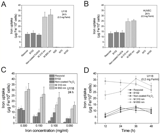
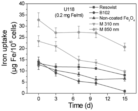
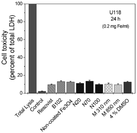
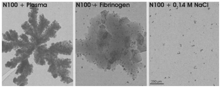
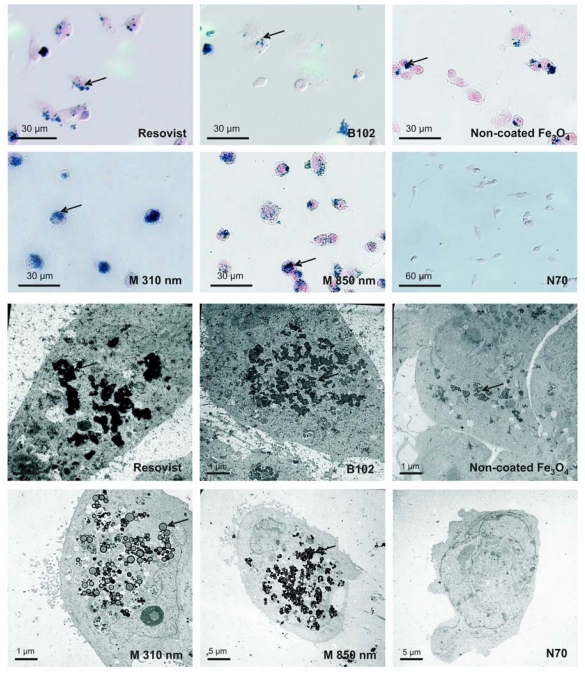
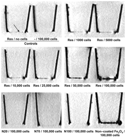
Superparamagnetic iron-oxide particles (SPIO) are used in different ways as contrast agents for magnetic resonance imaging (MRI): Particles with high nonspecific uptake are required for unspecific labeling of phagocytic cells whereas those that target specific molecules need to have very low unspecific cellular uptake. We compared iron-oxide particles with different core materials (magnetite, maghemite), different coatings (none, dextran, carboxydextran, polystyrene) and different hydrodynamic diameters (20-850 nm) for internalization kinetics, release of internalized particles, toxicity, localization of particles and ability to generate contrast in MRI. Particle uptake was investigated with U118 glioma cells und human umbilical vein endothelial cells (HUVEC), which exhibit different phagocytic properties. In both cell types, the contrast agents Resovist, B102, non-coated Fe(3)O(4) particles and microspheres were better internalized than dextran-coated Nanomag particles. SPIO uptake into the cells increased with particle/iron concentrations. Maximum intracellular accumulation of iron particles was observed between 24 h to 36 h of exposure. Most particles were retained in the cells for at least two weeks, were deeply internalized, and only few remained adsorbed at the cell surface. Internalized particles clustered in the cytosol of the cells. Furthermore, all particles showed a low toxicity. By MRI, monolayers consisting of 5000 Resovist-labeled cells could easily be visualized. Thus, for unspecific cell labeling, Resovist and microspheres show the highest potential, whereas Nanomag particles are promising contrast agents for target-specific labeling.
Keywords: cellular localization; electron microscopy; iron oxide nanoparticles; iron staining; magnetic resonance imaging (MRI); toxicity; uptake kinetics.
Figures
References
-
- Grimm J, Kircher M, Weissleder R. Cell tracking: Principles and applications. Radiologe. 2007;47:25–33. - PubMed
-
- Yoo BM, Pagel D. An overview of responsive MRI contrast agents for molecular imaging. Front. Biosci. 2008;13:1733–1752. - PubMed
-
- Nunn AD, Linder KE, Tweedle MF. Can receptors be imaged with MRI agents? Q. J. Nucl. Med. 1997;41:155–162. - PubMed
-
- Moore A, Weissleder R, Bogdanov A., Jr Uptake of dextran-coated monocrystalline iron oxides in tumor cells macrophages. J. Magn. Reson Imaging. 1997;7:1140–1145. - PubMed
Publication types
MeSH terms
Substances
LinkOut - more resources
Full Text Sources
Other Literature Sources
Medical
