

Construction and characterization of a cDNA library from wheat infected with Fusarium graminearum Fg 2
- PMID: 21340003
- PMCID: PMC3039969
- DOI: 10.3390/ijms12010613
Construction and characterization of a cDNA library from wheat infected with Fusarium graminearum Fg 2
Abstract
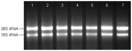
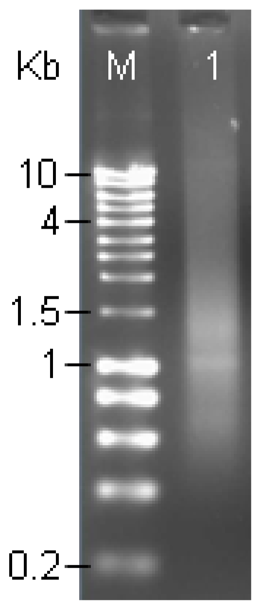
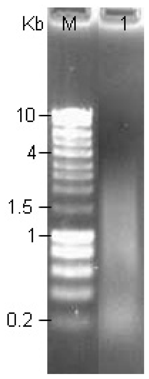
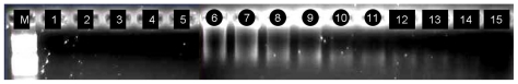
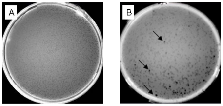
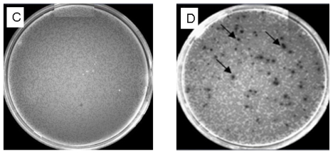
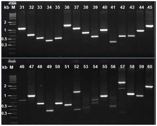
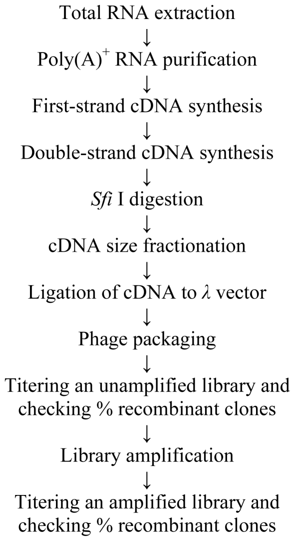
Total RNA from wheat spikes infected with F. graminearum Fg2 was extracted and the mRNA was purified. Switching Mechanism at 5' end of the RNA Transcript (SMART) technique and CDS Ill/3' primer were used for first-strand cDNA synthesis using reverse transcriptase by RT-PCR. Primer extension polymerase chain reaction was used to construct the double-strand cDNA that was digested by proteinase K, then by Sfi I and fractionated. cDNAs longer than 0.5 kb were collected and ligated to λTriplEx2 vector followed λ phage packaging reaction and library amplification. The qualities of both unamplified and amplified cDNA libraries were strictly checked by conventional titer determination. One hundred and sixty five plaques were randomly picked and tested using PCR with universal primers derived from the sequence flanking the vector. A high quality cDNA library from wheat spikes that have been infected by F. graminearum was successfully constructed.
Keywords: 15 acetyl deoxynivalenol (15ADON); 3 acetyl deoxynivalenol (3ADON); Fusarium graminearum Fg2; Triticum aestivum; cDNA library.
Figures
Similar articles
-
Transcriptome profiling of wheat differentially expressed genes exposed to different chemotypes of Fusarium graminearum.Theor Appl Genet. 2014 Aug;127(8):1703-18. doi: 10.1007/s00122-014-2333-8. Epub 2014 Jun 4. Theor Appl Genet. 2014. PMID: 24893796
-
Construction and characterization of a cDNA library from human liver tissue with chronic hepatitis B.J Zhejiang Univ Sci B. 2005 Apr;6(4):288-94. doi: 10.1631/jzus.2005.B0288. J Zhejiang Univ Sci B. 2005. PMID: 15754427 Free PMC article.
-
[Construction and characterization of a cDNA library from human liver tissue of cirrhosis].Zhejiang Da Xue Xue Bao Yi Xue Ban. 2005 Mar;34(2):98-103. doi: 10.3785/j.issn.1008-9292.2005.02.002. Zhejiang Da Xue Xue Bao Yi Xue Ban. 2005. PMID: 15812880 Chinese.
-
TRI12 based quantitative real-time PCR assays reveal the distribution of trichothecene genotypes of F. graminearum and F. culmorum isolates in Danish small grain cereals.Int J Food Microbiol. 2012 Jul 16;157(3):384-92. doi: 10.1016/j.ijfoodmicro.2012.06.010. Epub 2012 Jun 25. Int J Food Microbiol. 2012. PMID: 22781579
-
Transcriptomics of cereal-Fusarium graminearum interactions: what we have learned so far.Mol Plant Pathol. 2018 Mar;19(3):764-778. doi: 10.1111/mpp.12561. Epub 2017 Jun 7. Mol Plant Pathol. 2018. PMID: 28411402 Free PMC article. Review.
Cited by
-
Transcriptome profiling of wheat differentially expressed genes exposed to different chemotypes of Fusarium graminearum.Theor Appl Genet. 2014 Aug;127(8):1703-18. doi: 10.1007/s00122-014-2333-8. Epub 2014 Jun 4. Theor Appl Genet. 2014. PMID: 24893796
-
Construction of a full-length enriched cDNA library and preliminary analysis of expressed sequence tags from Bengal Tiger Panthera tigris tigris.Int J Mol Sci. 2013 May 24;14(6):11072-83. doi: 10.3390/ijms140611072. Int J Mol Sci. 2013. PMID: 23708105 Free PMC article.
-
Isolation and characterization of systemic acquired resistance marker gene PR1 and its promoter from Brassica juncea.3 Biotech. 2018 Jan;8(1):10. doi: 10.1007/s13205-017-1027-8. Epub 2017 Dec 11. 3 Biotech. 2018. PMID: 29259885 Free PMC article.
References
-
- Bai GH, Shaner G. Scab of wheat: Prospects for control. Plant Dis. 1994;78:760–766.
-
- Diatchenko L, Lar YFC, Campbell AP, Chenchik A, Moqadam F, Huang B, Lukyanov S, Lukyanov K, Gurskaya N, Sverdlov ED, Silbert PD. Suppression subtractive hybridization: A method for generating differentially regulated or tissue-specific cDNA probes and libraries. Proc. Natl. Acad. Sci USA. 1996;93:6025–6030. - PMC - PubMed
-
- Wang X, Feuerstein GZ. The use of mRNA differential display for discovery of novel therapeutic targets in cardiovascular disease. Cardiovasc. Res. 1997;35:414–421. - PubMed
-
- Sambrook J, Russell DW. Molecular Cloning: A Laboratory Manual. 3rd ed. Cold Spring Harbor Lab Press; New York, NY, USA: 2002.
-
- Draper MP, August PR, Connolly T, Packard B, Call KM. Efficient cloning of full-length cDNAs based on cDNA size fractionation. Genomics. 2002;79:603–607. - PubMed
Publication types
MeSH terms
LinkOut - more resources
Full Text Sources
