. 2011 Feb 14;6(2):e17042.
doi: 10.1371/journal.pone.0017042.

Genetically engineered alginate lyase-PEG conjugates exhibit enhanced catalytic function and reduced immunoreactivity

Affiliations

Genetically engineered alginate lyase-PEG conjugates exhibit enhanced catalytic function and reduced immunoreactivity

John W Lamppa et al. PLoS One. .

Abstract

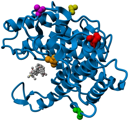
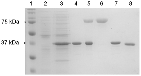
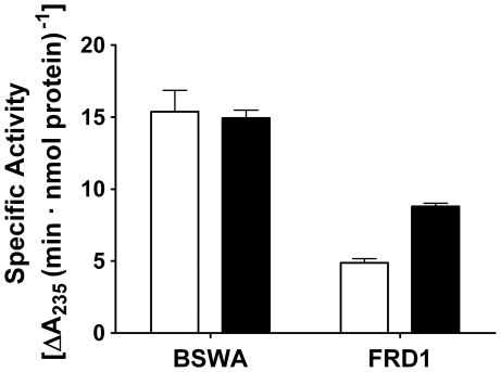
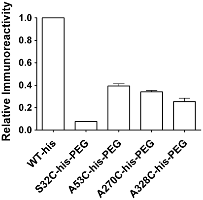
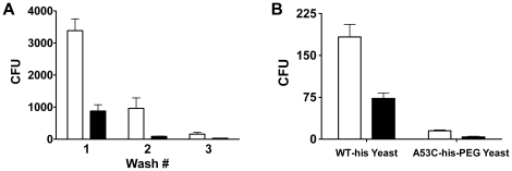
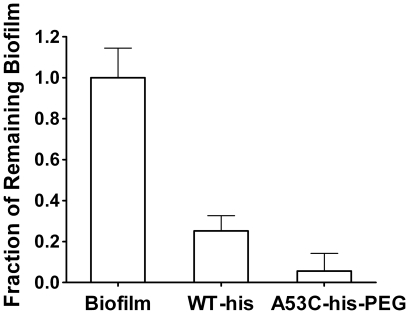
Alginate lyase enzymes represent prospective biotherapeutic agents for treating bacterial infections, particularly in the cystic fibrosis airway. To effectively deimmunize one therapeutic candidate while maintaining high level catalytic proficiency, a combined genetic engineering-PEGylation strategy was implemented. Rationally designed, site-specific PEGylation variants were constructed by orthogonal maleimide-thiol coupling chemistry. In contrast to random PEGylation of the enzyme by NHS-ester mediated chemistry, controlled mono-PEGylation of A1-III alginate lyase produced a conjugate that maintained wild type levels of activity towards a model substrate. Significantly, the PEGylated variant exhibited enhanced solution phase kinetics with bacterial alginate, the ultimate therapeutic target. The immunoreactivity of the PEGylated enzyme was compared to a wild type control using in vitro binding studies with both enzyme-specific antibodies, from immunized New Zealand white rabbits, and a single chain antibody library, derived from a human volunteer. In both cases, the PEGylated enzyme was found to be substantially less immunoreactive. Underscoring the enzyme's potential for practical utility, >90% of adherent, mucoid, Pseudomonas aeruginosa biofilms were removed from abiotic surfaces following a one hour treatment with the PEGylated variant, whereas the wild type enzyme removed only 75% of biofilms in parallel studies. In aggregate, these results demonstrate that site-specific mono-PEGylation of genetically engineered A1-III alginate lyase yielded an enzyme with enhanced performance relative to therapeutically relevant metrics.


Conflict of interest statement

Competing Interests: The authors have declared that no competing interests exist.

Figures

Figure 1
Figure 1. Sites of cysteine substitution.
Ribbon diagram of alginate Lyase A1-III (PDB file 1HV6). A trisaccharide reaction product is bound in the active cleft and shown as a grey ball and stick model. Amino acid residues targeted for cysteine substitution are shown in space filling mode, and are color coded as follows: S32C = Red, A41C = Orange, A53C = Green, A270C = Yellow, and A328C = Purple.
Figure 2
Figure 2. SDS-PAGE analysis of PEGylated variant production.
Samples run on a reducing 12.5% gel, and stained for total protein with Coomassie brilliant blue. Lane 1: Bio-Rad Precision Plus Protein Ladder; Lane 2: Whole cell lysate of non-expressing cells; Lane 3: Whole cell lysate of induced cells; Lane 4: IMAC purified A53C-his; Lane 5: Crude A53C-his PEGylation reaction product; Lane 6: Size exclusion FPLC purified A53C-his-PEG; Lane 7: IMAC purified WT-his; Lane 8: FPLC purified native WT.
Figure 3
Figure 3. Comparison of reaction kinetics with BSWA and bacterial alginate.
The specific activities of WT-his (white bars) and A53C-his-PEG (black bars) were determined with a model alginate substrate (BSWA) as well as with purified bacterial alginate (FRD1). The two enzymes are equally active with BSWA at saturating concentrations, but the PEGylated variant exhibits 80% faster kinetics with the bacterial substrate (p<0.01), which is the ultimate therapeutic target. Error bars represent standard deviation.
Figure 4
Figure 4. Immunoreactivity by ELISA.
The antibody concentration required to achieve 50% maximum ELISA signal (EC50) was determined for each enzyme using polyclonal anti-A1-III antibody purified from rabbit immune serum. The results are reported as fractional immunoreactivity based on normalization with the WT-his enzyme, which was included as an internal control in all experiments (see Experimental Procedures). All of the PEGylated enzymes were found to exhibit significantly reduced antibody binding relative to the WT-his control (p<0.01 for each). Error bars represent standard deviation.
Figure 5
Figure 5. Human antibody binding.
The WT-his (white bars) and A53C-his-PEG (black bars) protein targets were biotinylated and captured on the surface of streptavidin coated magnetic beads. A) The two bead preparations were independently incubated with a yeast surface displayed scFv antibody library derived from human immune cells. Following binding, the beads were magnetically separated and washed three times. The number of yeast that remained bound after each wash step was determined by plating serial dilutions of the resuspended beads and enumerating cfu's. The resulting yeast colonies represent human scFvs that specifically bound to the A1-III enzymes on the corresponding magnetic beads. A53C-his-PEG coated magnetic beads bound up to 13-fold fewer human antibodies than did the WT-his coated beads (p<0.01 for each of the three washes). B) Characterization of first round binders from both protein targets. Yeast isolated as binders to either WT-his or A53C-his-PEG were propagated and subsequently incubated with magnetic beads bearing each protein target. For both yeast populations, the A53C-his-PEG coated beads (black bars) bound at least 60% fewer cells than did the WT-his beads (white bars), a result that demonstrates PEGylation effectively blocked key immunogenic epitopes (p<0.01 for all differences). Error bars represent standard deviation.
Figure 6
Figure 6. Disruption of mucoid P. aeruginosa biofilms.
Adherent biofilms of a mucoid clinical isolate were established in 96-well plates and subsequently treated with 1 mg/ml enzyme for 1 hour. Remaining biofilm was then quantified using an alginate-sensitive lectin-HRP conjugate and ABTS substrate. Signals were normalized to a buffer only treatment. Both enzymes removed a significant proportion of biofilm relative to the buffer control (p<0.01). Importantly, theA53C-his-PEG enzyme removed >15% more biofilm than the WT-his enzyme (p<0.025). Error bars represent standard deviation.

References

    1. Elkin S, Geddes D. Pseudomonal infection in cystic fibrosis: the battle continues. Expert Review of Anti-Infective Therapy. 2003;1:609–618. - PubMed
    1. May TB, Shinabarger D, Maharaj R, Kato J, Chu L, et al. Alginate synthesis by Pseudomonas aeruginosa: a key pathogenic factor in chronic pulmonary infections of cystic fibrosis patients. Clinical Microbiology Reviews. 1991;4:191–206. - PMC - PubMed
    1. Mai GT, Seow WK, Pier GB, McCormack JG, Thong YH. Suppression of Lymphocyte and neutrophil Functions by Pseudomonas aeruginosa mucoid exopolysaccharide (alginate): Reversal by physicochemical, alginase, and specific monoclonal antibody treatments. Infection and Immunity. 1993;61:559–564. - PMC - PubMed
    1. Simpson JA, Smith SE, Dean RT. Alginate may accumulate in cystic fibrosis lung because the enzymatic and free radical capacities of phagocytic cells are inadequate for its degradation. Biochemistry and Molecular Biology International. 1993;6:1021–1034. - PubMed
    1. Hoiby N, Krogh Johansen H, Moser C, Song Z, Ciofu O, et al. Pseudomonas aeruginosa and the in vitro and in vivo biofilm mode of growth. Microbes and Infection. 2001;3:23–35. - PubMed

Publication types