Review
. 2011 Apr;3(4):279-96.
doi: 10.1039/c0ib00144a. Epub 2011 Feb 22.

Apoptotic cell signaling in cancer progression and therapy

Affiliations
Review

Apoptotic cell signaling in cancer progression and therapy

Jessica Plati et al. Integr Biol (Camb). 2011 Apr.

Abstract

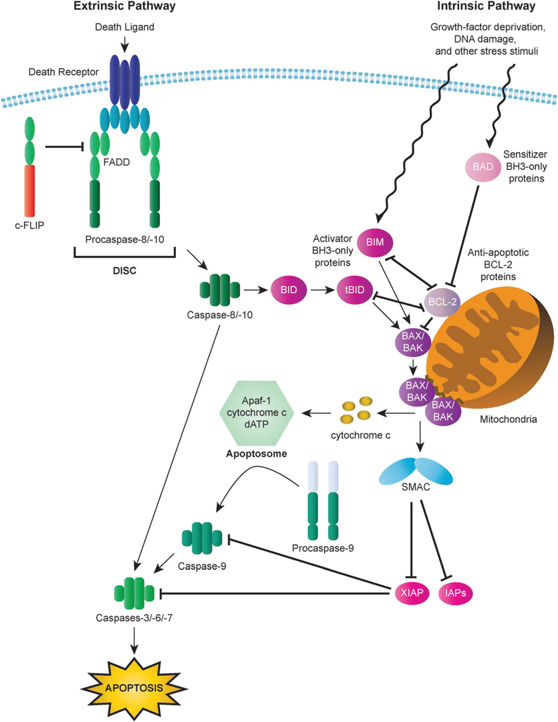
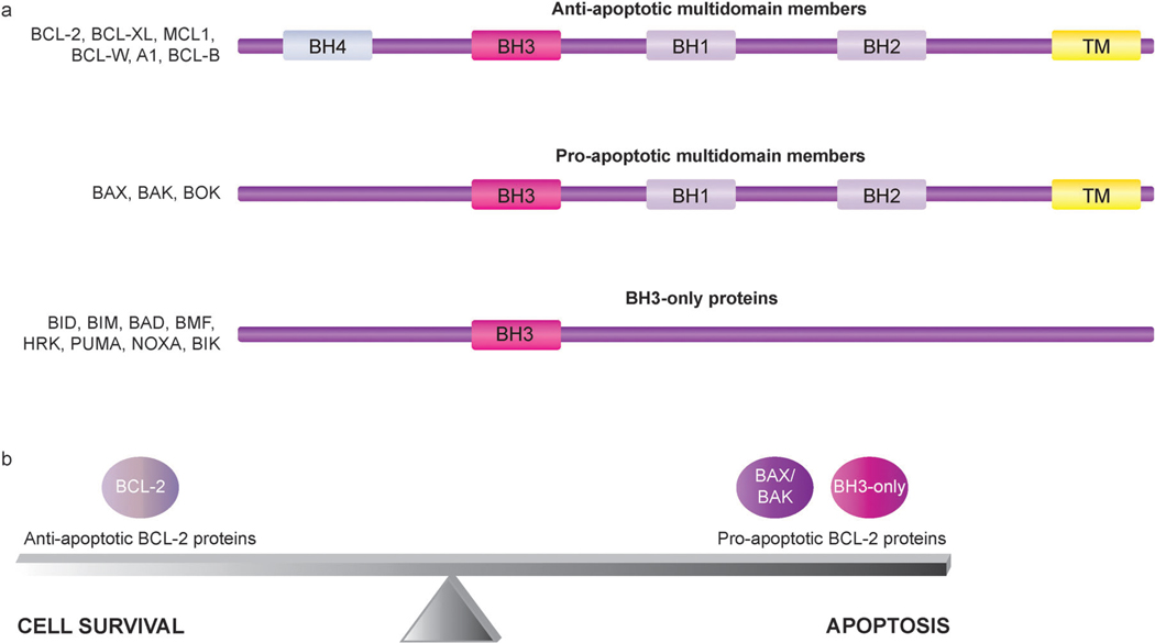
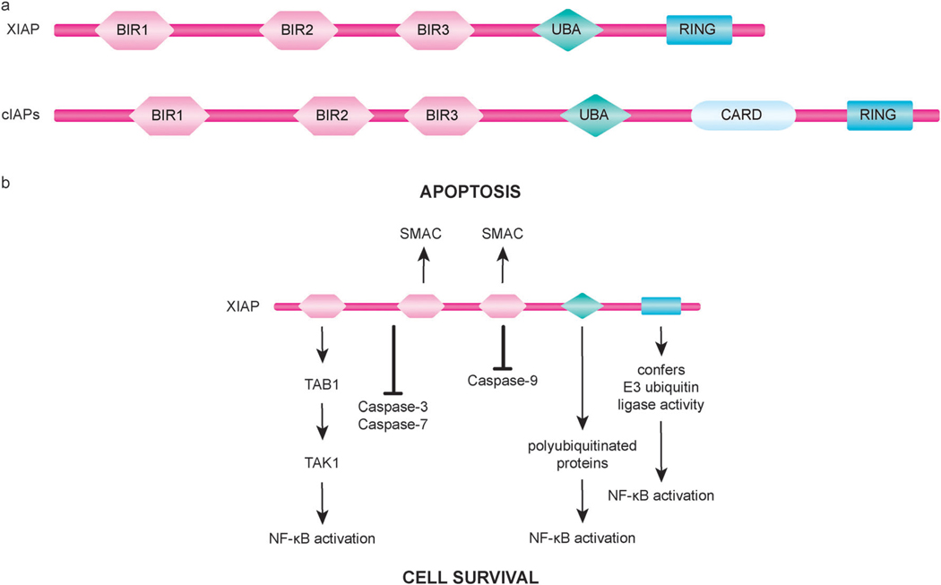
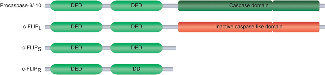
Apoptosis is a tightly regulated cell suicide program that plays an essential role in the development and maintenance of tissue homeostasis by eliminating unnecessary or harmful cells. Impairment of this native defense mechanism promotes aberrant cellular proliferation and the accumulation of genetic defects, ultimately resulting in tumorigenesis, and frequently confers drug resistance to cancer cells. The regulation of apoptosis at several levels is essential to maintain the delicate balance between cellular survival and death signaling that is required to prevent disease. Complex networks of signaling pathways act to promote or inhibit apoptosis in response to various cues. Apoptosis can be triggered by signals from within the cell, such as genotoxic stress, or by extrinsic signals, such as the binding of ligands to cell surface death receptors. Various upstream signaling pathways can modulate apoptosis by converging on, and thereby altering the activity of, common central control points within the apoptotic signaling pathways, which involve the BCL-2 family proteins, inhibitor of apoptosis (IAP) proteins, and FLICE-inhibitory protein (c-FLIP). This review highlights the role of these fundamental regulators of apoptosis in the context of both normal apoptotic signaling mechanisms and dysregulated apoptotic pathways that can render cancer cells resistant to cell death. In addition, therapeutic strategies aimed at modulating the activity of BCL-2 family proteins, IAPs, and c-FLIP for the targeted induction of apoptosis are briefly discussed.


Figures

Fig. 1
Fig. 1
Intrinsic and extrinsic apoptotic pathways. In the intrinsic (mitochondrial) pathway, mitochondrial outer membrane permeabilization (MOMP) results in the release of cytochrome c and other apoptogenic factors from mitochondria into the cytosol and the ensuing formation of the apoptosome, which triggers activation of the apoptosis-inducing caspase cascade via activation of caspase-9. Interactions among BCL-2 family proteins play a critical role in the mediating MOMP induction and consequent apoptosis. BH3-only proteins (activator BH3-only proteins are represented here by BIM and tBID and sensitizer BH3-only proteins are represented by BAD) can relay apoptotic signals to the mitochondria through activation of BAX or BAK, the principal effectors of the intrinsic apoptotic pathway. In contrast, anti-apoptotic BCL-2 proteins (represented by BCL-2) serve to inhibit apoptosis by blocking BAX/BAK activation. In the extrinsic (death receptor) pathway, binding of death receptors such as FAS or TRAIL receptors (TRAILR1, TRAILR2) by their cognate ligands triggers the recruitment of death domain (DD)-containing adaptor proteins (represented by FADD) and procaspases with a death effector domain (DED), specifically procaspase-8 and procaspase-10. The resulting complex is known as the death inducing signaling complex (DISC). High levels of active caspase-8 generated by large amounts of procaspase-8 processing at the DISC lead to the activation of executioner caspases, including caspase-3, and the induction of apoptosis. Activation of caspase-8 can also result in the cleavage of the BH3-only protein BID to generate the activated BID fragment tBID, which serves to transmit the death signal from the extrinsic to the intrinsic signaling pathway.
Fig. 2
Fig. 2
Subgroups of BCL-2 family members with representative members of each subfamily. (a) BCL-2 family members can be classified into three subgroups according to function and BH domain composition. All BCL-2 family members possess at least one of four BCL-2 homology (BH) domains, termed BH1, BH2, BH3, and BH4, and many also include a transmembrane (TM) domain. The anti-apoptotic multidomain members have three to four BH domains, with some members lacking a BH4 domain. Similar to the anti-apoptotic multidomain members, the pro-apoptotic multidomain members contain BH1, BH2, and BH3 domains. The BH3-only proteins are a subset of pro-apoptotic proteins that only bear a single BH motif, the BH3 domain. Some BH3-only proteins also include a TM domain. (b) BCL-2 proteins play a key role in mediating the delicate balance between cell survival and cell death. Disruption of this balance by cellular alterations that increase the functional activity of anti-apoptotic BCL-2 proteins relative to pro-apoptotic BCL-2 proteins can enable the evasion of apoptosis, which tips the balance to favor cell survival and thus promotes the development and progression of cancer.
Fig. 3
Fig. 3
Domain organization and function of inhibitors of apoptosis (IAP) proteins. (a) XIAP, a well studied human IAP family member, and the structurally similar family members cIAP1 and cIAP2 (cIAPs) each have three tandem BIR domains followed by an ubiquitin-associated (UBA) domain and a C-terminal RING finger domain. cIAPs also possess a caspase recruitment domain (CARD) of unknown function located between the UBA and the RING domains. (b) The BIR2 domain of XIAP, along with residues in its N-terminal flanking linker region, mediates the binding and inhibition of caspase-3 and caspase-7. Inactivation of caspase-9 by XIAP involves the BIR3 domain of XIAP binding to caspase-9. In addition to blocking caspase activity, XIAP can also promote cell survival through regulation of important cellular signaling pathways, including signaling mechanisms of NF-κB activation. IAP-binding motif (IBM)-containing proteins, such as SMAC, interact with the BIR2 and BIR3 domains of XIAP to neutralize its anti-apoptotic activity.
Fig. 4
Fig. 4
Regulation of NF-κB signaling by cIAP1 and cIAP2 (cIAPs). cIAPs play a key role in TNF-induced NF-κB activation by functioning as E3 ligases that ubiquitinate RIP1, leading to the stabilization of complex I and activation of NF-κB. In the presence of SMAC mimetics, cIAPs undergo auto-ubiquitination and proteasomal degradation. The loss of cIAPs sensitizes cells to apoptosis in response to TNF by allowing the formation of complex II, which requires deubiquitination of RIP1. Complex II contains RIP1, FADD, and procaspase-8 and serves to trigger apoptosis by promoting activation of caspase-8. Interestingly, cIAPs can also act as negative regulators of NF-κB activation by targeting NF-κB-inducing kinase (NIK) for proteasomal degradation. In this context, the loss of cIAPs induced by SMAC mimetics promotes NIK stabilization and consequent NF-κB activation.
Fig. 5
Fig. 5
Structural features of the FLICE-inhibitory protein (c-FLIP) isoforms. Each of the three c-FLIP isoforms, c-FLIPL, c-FLIPS, and c-FLIPR, contain two tandem death effector domains (DEDs) at its N-terminus. c-FLIPL, the long c-FLIP isoform, is similar in overall structure to procaspase-8, but its caspase-like domain is functionally inactive. Other than the two DEDs, the short c-FLIP isoforms c-FLIPS and c-FLIPR only include a short C-terminus.

References

    1. Cotter TG. Apoptosis and cancer: the genesis of a research field. Nat. Rev. Cancer. 2009;9:501–507. - PubMed
    1. Kerr JF, Wyllie AH, Currie AR. Apoptosis: a basic biological phenomenon with wide-ranging implications in tissue kinetics. Br. J. Cancer. 1972;26:239–257. - PMC - PubMed
    1. Hanahan D, Weinberg RA. The hallmarks of cancer. Cell. 2000;100:57–70. - PubMed
    1. Halazonetis TD, Gorgoulis VG, Bartek J. An oncogene-induced DNA damage model for cancer development. Science. 2008;319:1352–1355. - PubMed
    1. Negrini S, Gorgoulis VG, Halazonetis TD. Genomic instability-an evolving hallmark of cancer. Nat. Rev. Mol. Cell Biol. 2010;11:220–228. - PubMed

Publication types