

[Endoscopic ligation of the anterior ethmoidal artery: a cadaver dissection study]
- PMID: 21340186
- PMCID: PMC9442171
- DOI: 10.1590/s1808-86942011000100006
[Endoscopic ligation of the anterior ethmoidal artery: a cadaver dissection study]
Abstract
Anterior ethmoidal artery (AEA) ligation may be necessary in cases of severe epistaxis not controllable with traditional therapy. Endoscopic endonasal ligation of the AEA is not used frequently; there are few studies in the literature for standardization of the endoscopic technique for this vessel.
Aim: To demonstrate the feasibility of periorbital AEA ligation in a transethmoidal endoscopic approach.
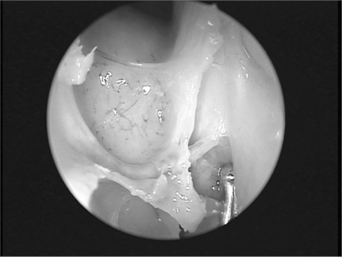
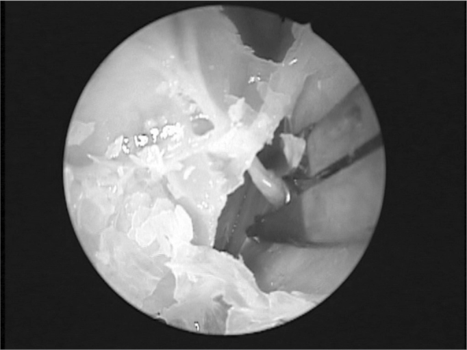
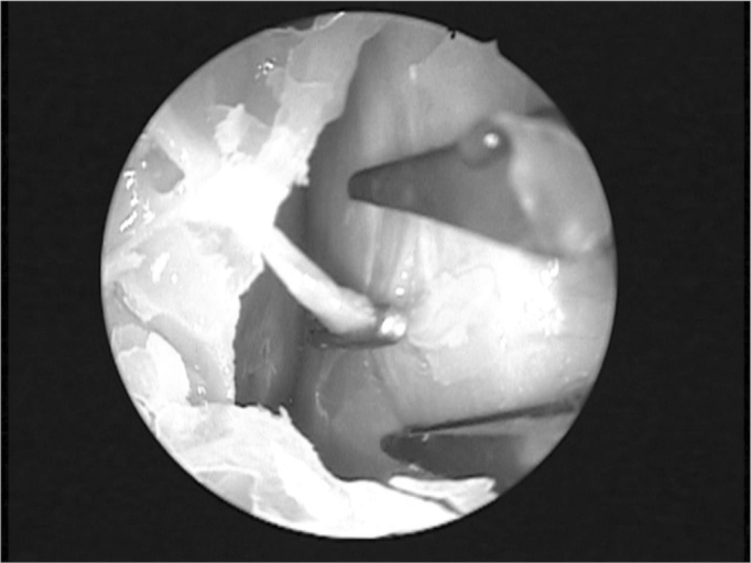
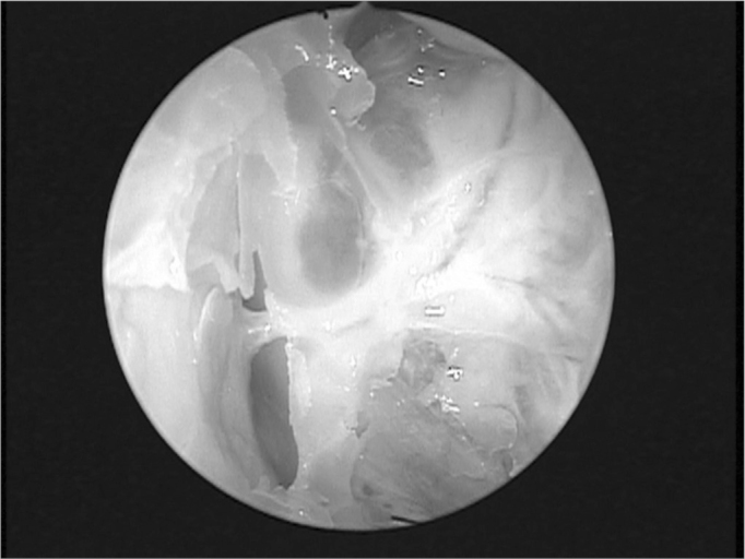
Methods: A prospective study where 50 nasal cavities were dissected. After anterior ethmoidectomy and partial removal of lamina papyracea, the periorbital area was carefully dissected along a subperiosteal plane to identify the AEA. The vessel was exposed within the orbit and dissected.
Results: Data on technical difficulties, complications, the learning curve and anatomical variations were gathered.
Conclusion: An endonasal endoscopic approach to the AEA within the orbit was shown to be feasible. Identifying the artery is not difficult, and this technique avoids external incisions. This approach appears to be an excellent alternative for approaching the AEA. Further clinical studies are needed to demonstarte the benefits of this technique.
Figures
References
-
- Rockey JG, Anand R. A critical audit of the surgical management of intractable epistaxis using sphenopalatine artery ligation/diathermy. Rhinology. 2002;40:147–149. - PubMed
-
- Douglas SA, Gupta D. Endoscopic assisted esternal approach anterior ethmoidal artery ligation for management of epistaxis. J Laryngol Otol. 2003;117:132–133. - PubMed
-
- Voegels RL. Cirurgia endoscopica dos seios paranasais. Arq Int Otorrinolaringol. 1997;1(1):15–18.
-
- Busch RF. A new vascular clip applier for internal maxillary and ethmoidal artery ligations. Otolaryngol Head Neck Surg. 1992;107(1):129–130. - PubMed
-
- Singh B. Combined Internal maxillary and Anterior Ethmoidal artery occlusion: The treatment of choice in intractable epistaxis. J Laryngol Otol. 1992;106:507–510. - PubMed
Publication types
MeSH terms
LinkOut - more resources
Full Text Sources
Medical
