

Pathological and ultrastructural analysis of surgical lung biopsies in patients with swine-origin influenza type A/H1N1 and acute respiratory failure
- PMID: 21340209
- PMCID: PMC3020331
- DOI: 10.1590/s1807-59322010001200003
Pathological and ultrastructural analysis of surgical lung biopsies in patients with swine-origin influenza type A/H1N1 and acute respiratory failure
Abstract
Background: Cases of H1N1 and other pulmonary infections evolve to acute respiratory failure and death when co-infections or lung injury predominate over the immune response, thus requiring early diagnosis to improve treatment.
Objective: To perform a detailed histopathological analysis of the open lung biopsy specimens from five patients with ARDS with confirmed H1N1.
Methods: Lung specimens underwent microbiologic analysis, and examination by optical and electron microscopy. Immunophenotyping was used to characterize macrophages, natural killer, T and B cells, and expression of cytokines and iNOS.
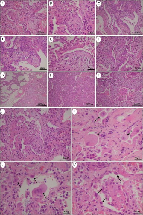
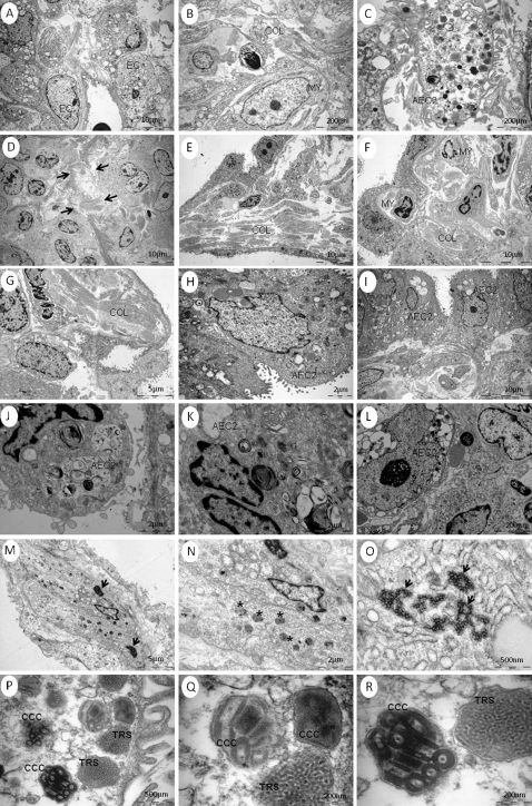
Results: The pathological features observed were necrotizing bronchiolitis, diffuse alveolar damage, alveolar hemorrhage and abnormal immune response. Ultrastructural analysis showed viral-like particles in all cases.
Conclusions: Viral-like particles can be successfully demonstrated in lung tissue by ultrastructural examination, without confirmation of the virus by RT-PCR on nasopharyngeal aspirates. Bronchioles and epithelium, rather than endothelium, are probably the primary target of infection, and diffuse alveolar damage the consequence of the effect of airways obliteration and dysfunction on innate immunity, suggesting that treatment should be focused on epithelial repair.
Figures

References
-
- Centers for Disease Control and Prevention (CDC) Swine influenza 1. A (H1N1) infection in two children — Southern California, March–April 2009. MMWR Morb Mortal Wkly Rep. 2009;58:400–2. - PubMed
-
- Centers for Disease Control and Prevention (CDC) Update: infections with a swine‐origin influenza A (H1N1) virus — United States and other countries, April 28, 2009. MMWR Morb Mortal Wkly Rep. 2009;58:431–3. - PubMed
-
- Centers for Disease Control and Prevention (CDC) Update: swine influenza A (H1N1) infections — California and Texas, April 2009. MMWR Morb Mortal Wkly Rep. 2009;58:435–7. - PubMed
-
- Novel Swine‐Origin Influenza A (H1N1) Virus Investigation Team. Emergence of a novel swine‐origin influenza A (H1N1) virus in humans. N Engl J Med. 2009;360:2605–15. 10.1056/NEJMoa0903810 - DOI - PubMed
-
- World Health Organization ‐ pandemic (H1N1) 2009 ‐ update 77. 2009 December 4. Available from: http://www.who.int/csr/don/2009_10_09/en/
Publication types
MeSH terms
LinkOut - more resources
Full Text Sources
Medical
