. 2011 Feb 22:8:76.
doi: 10.1186/1743-422X-8-76.

Peptide inhibition of human cytomegalovirus infection

Affiliations

Peptide inhibition of human cytomegalovirus infection

Lilia I Melnik et al. Virol J. .

Abstract

Background: Human cytomegalovirus (HCMV) is the most prevalent congenital viral infection in the United States and Europe causing significant morbidity and mortality to both mother and child. HCMV is also an opportunistic pathogen in immunocompromised individuals, including human immunodeficiency virus (HIV)- infected patients with AIDS, and solid organ and allogeneic stem cell transplantation recipients. Current treatments for HCMV-associated diseases are insufficient due to the emergence of drug-induced resistance and cytotoxicity, necessitating novel approaches to limit HCMV infection. The aim of this study was to develop therapeutic peptides targeting glycoprotein B (gB), a major glycoprotein of HCMV that is highly conserved across the Herpesviridae family, that specifically inhibit fusion of the viral envelope with the host cell membrane preventing HCMV entry and infection.

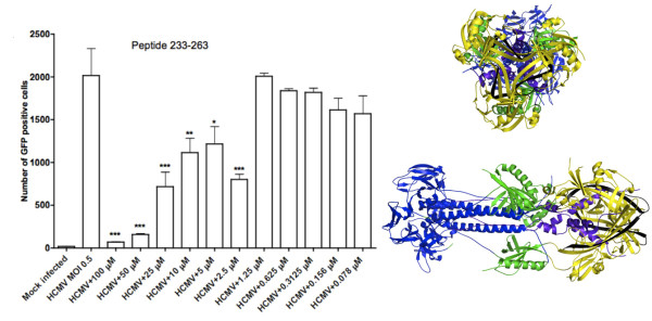
Results: Using the Wimley-White Interfacial Hydrophobicity Scale (WWIHS), several regions within gB were identified that display a high potential to interact with lipid bilayers of cell membranes and hydrophobic surfaces within proteins. The ability of synthetic peptides analogous to WWIHS-positive sequences of HCMV gB to inhibit viral infectivity was evaluated. Human foreskin fibroblasts (HFF) were infected with the Towne-GFP strain of HCMV (0.5 MOI), preincubated with peptides at a range of concentrations (78 nm to 100 μM), and GFP-positive cells were visualized 48 hours post-infection by fluorescence microscopy and analyzed quantitatively by flow cytometry. Peptides that inhibited HCMV infection demonstrated different inhibitory concentration curves indicating that each peptide possesses distinct biophysical properties. Peptide 174-200 showed 80% inhibition of viral infection at a concentration of 100 μM, and 51% and 62% inhibition at concentrations of 5 μM and 2.5 μM, respectively. Peptide 233-263 inhibited infection by 97% and 92% at concentrations of 100 μM and 50 μM, respectively, and 60% at a concentration of 2.5 μM. While peptides 264-291 and 297-315, individually failed to inhibit viral infection, when combined, they showed 67% inhibition of HCMV infection at a concentration of 0.125 μM each.

Conclusions: Peptides designed to target putative fusogenic domains of gB provide a basis for the development of novel therapeutics that prevent HCMV infection.


Figures

Figure 1
Figure 1
Wimley-White interfacial hydrophobicity scale score-positive sequences in class II and III viral fusion proteins. Sequences of a representative class II viral fusion protein (Dengue virus E) and of class III viral fusion proteins with high potential to interface with lipid membranes (red) were identified using Membrane Protein Explorer software (MpeX version 3.0). As discussed in the text, a class III domain nomenclature is used here that can apply to both class II and III viral fusions proteins. The alternative domain number schemes used by Roche et al. and Heldwein et al. are noted in parentheses. The Dengue virus (DENV E) stem domain sequence that was not included in the protein used to determine the crystal structure has been added. The DENV stem has a positive WWIHS scale score, and corresponds to a previously determined inhibitor of DENV and West Nile virus [35].
Figure 2
Figure 2
Determination of regions within gB that display a high propensity to interact with the lipid surface of cell membranes by using Wimley-White Interfacial Hydrophobicity Scale (WWIHS). WWIHS identifies segments of proteins that prefer a transbilayer helix conformation to an unfolded interfacial location. We used the Interface Scale of the Membrane Protein explorer (MpeX version 3.0) computer program to identify these particular segments of HCMV gB. The Interface scale measures a residue's free energy of transfer within an unfolded polypeptide chain from water to a phosphocholine bilayer. We identified nine segments of HCMV gB that display high propensity to interact with the lipid surface of cell membrane, and designed peptides, ranging from 19 to 31 amino acids in length, that are analogous to the identified regions of gB.
Figure 3
Figure 3
Inhibition of HCMV infection by peptide 174-200. HFF were seeded at a density of 3.5×10⁵ cells in each well of a 24-well plate 24 hours prior to infection. The inhibitory effect of peptide 174-200 was evaluated by infecting human foreskin fibroblasts (HFF) with the Towne-GFP strain of HCMV (0.5 MOI) preincubated with different concentrations of inhibitory peptide 174-200 at 37°C for 90 minutes. GFP-positive cells were visualized 48 hours post-infection by fluorescence microscopy and then quantified using flow cytometry. Significant reductions in the number of GFP-positive cells compared to HCMV infected cells are denoted by a * (p < 0.05), ** (p < 0.01), and *** (p < 0.001, determined using one-way ANOVA and Tukey's post test). All structural figures of HSV-1 gB in the post-fusion configuration were generated using MacPyMOL [56] and FreeHand (Macromedia). Different domains of gB are shown in the ribbon structures: yellow-fusion domain II, purple-stem, green-domain I, and blue-domain III with extended α-helices, which are involved in trimerization. Peptides targeting different domains of HSV-1 gB that correspond to HCMV gB domains are shown in black.
Figure 4
Figure 4
Inhibition of HCMV infection by peptide 233-263. HFF were seeded at a density of 3.5×10⁵ cells in each well of a 24-well plate 24 hours prior to infection. The inhibitory effect of peptide 233-263 was evaluated by infecting human foreskin fibroblasts (HFF) with the Towne-GFP strain of HCMV (0.5 MOI) preincubated with different concentrations of inhibitory peptide 233-263 at 37°C for 90 minutes. GFP-positive cells were visualized 48 hours post-infection by fluorescence microscopy and then quantified using flow cytometry. Significant reductions in the number of GFP-positive cells compared to HCMV infected cells are denoted by a * (p < 0.05), ** (p < 0.01), and *** (p < 0.001, determined using one-way ANOVA and Tukey's post test). All structural figures of HSV-1 gB in the post-fusion configuration were generated using MacPyMOL [56] and FreeHand (Macromedia). Different domains of gB are shown in the ribbon structures: yellow-fusion domain II, purple-stem, green-domain I, and blue-domain III with extended α-helices, which are involved in trimerization. Peptides targeting different domains of HSV-1 gB that correspond to HCMV gB domains are shown in black.
Figure 5
Figure 5
Inhibition of HCMV infection by peptide 264-291. HFF were seeded at a density of 3.5×10⁵ cells in each well of a 24-well plate 24 hours prior to infection. The inhibitory effect of peptide 264-291 was evaluated by infecting human foreskin fibroblasts (HFF) with the Towne-GFP strain of HCMV (0.5 MOI) preincubated with different concentrations of inhibitory peptide 264-291 at 37°C for 90 minutes. GFP-positive cells were visualized 48 hours post-infection by fluorescence microscopy and then quantified using flow cytometry. Significant reductions in the number of GFP-positive cells compared to HCMV infected cells are denoted by a * (p < 0.05), ** (p < 0.01), and *** (p < 0.001, determined using one-way ANOVA and Tukey's post test). All structural figures of HSV-1 gB in the post-fusion configuration were generated using MacPyMOL [56] and FreeHand (Macromedia). Different domains of gB are shown in the ribbon structures: yellow-fusion domain II, purple-stem, green-domain I, and blue-domain III with extended α-helices, which are involved in trimerization. Peptides targeting different domains of HSV-1 gB that correspond to HCMV gB domains are shown in black.
Figure 6
Figure 6
Inhibition of HCMV infection by peptide 297-315. HFF were seeded at a density of 3.5×10⁵ cells in each well of a 24-well plate 24 hours prior to infection. The inhibitory effect of peptide 297-315 was evaluated by infecting human foreskin fibroblasts (HFF) with the Towne-GFP strain of HCMV (0.5 MOI) preincubated with different concentrations of inhibitory peptide 297-315 at 37°C for 90 minutes. GFP-positive cells were visualized 48 hours post-infection by fluorescence microscopy and then quantified using flow cytometry. Significant reductions in the number of GFP-positive cells compared to HCMV infected cells are denoted by a * (p < 0.05), ** (p < 0.01), and *** (p < 0.001, determined using one-way ANOVA and Tukey's post test). All structural figures of HSV-1 gB in the post-fusion configuration were generated using MacPyMOL [56] and FreeHand (Macromedia). Different domains of gB are shown in the ribbon structures: yellow-fusion domain II, purple-stem, green-domain I, and blue-domain III with extended α-helices, which are involved in trimerization. Peptides targeting different domains of HSV-1 gB that correspond to HCMV gB domains are shown in black.
Figure 7
Figure 7
Additive effect of HCMV gB peptides 174-200 and 233-263 on HCMV infection. HFF were seeded at a density of 3.5×10⁵ cells in each well of a 24-well plate 24 hours prior to infection. The ability of peptides 174-200 and 233-263 to work together and their effect on inhibition of virus infection was evaluated by infecting human foreskin fibroblasts (HFF) with the Towne-GFP strain of HCMV (0.5 MOI) preincubated with different concentrations of inhibitory peptides at 37°C for 90 minutes. GFP-positive cells were visualized 48 hours post-infection by fluorescence microscopy and then quantified using flow cytometry. Significant reductions in the number of GFP-positive cells compared to HCMV infected cells are denoted by a * (p < 0.05), ** (p < 0.01), and *** (p < 0.001, determined using one-way ANOVA and Tukey's post test). All structural figures of HSV-1 gB in the post-fusion configuration were generated using MacPyMOL [56] and FreeHand (Macromedia). Different domains of gB are shown in the ribbon structures: yellow-fusion domain II, purple-stem, green-domain I, and blue-domain III with extended α-helices, which are involved in trimerization. Peptides targeting different domains of HSV-1 gB that correspond to HCMV gB domains are shown in black.
Figure 8
Figure 8
Additive effect of HCMV gB peptides 264-291 and 297-315 on HCMV infection. HFF were seeded at a density of 3.5×10⁵ cells in each well of a 24-well plate 24 hours prior to infection. The ability of peptides 264-291 and 297-315 to work together and their effect on inhibition of virus infection was evaluated by infecting human foreskin fibroblasts (HFF) with the Towne-GFP strain of HCMV (0.5 MOI) preincubated with different concentrations of inhibitory peptides at 37°C for 90 minutes. GFP-positive cells were visualized 48 hours post-infection by fluorescence microscopy and then quantified using flow cytometry. Significant reductions in the number of GFP-positive cells compared to HCMV infected cells are denoted by a * (p < 0.05), ** (p < 0.01), and *** (p < 0.001, determined using one-way ANOVA and Tukey's post test). All structural figures of HSV-1 gB in the post-fusion configuration were generated using MacPyMOL [56] and FreeHand (Macromedia). Different domains of gB are shown in the ribbon structures: yellow-fusion domain II, purple-stem, green-domain I, and blue-domain III with extended α-helices, which are involved in trimerization. Peptides targeting different domains of HSV-1 gB that correspond to HCMV gB domains are shown in black.
Figure 9
Figure 9
Bright light and fluorescent images representing the ability of peptide 233-263 to inhibit infection by the Towne-GFP strain of HCMV. (A-B) Representative fluorescent and bright light microscopic images of HFF infected with the Towne-GFP strain of HCMV (0.5 MOI). (C-D). Representative fluorescent and bright light images of HFF infected with the Towne-GFP strain of HCMV (0.5 MOI) preincubated with peptide 233-263 at a concentration of 100 μM.
Figure 10
Figure 10
Comparison of HSV-1 gB-1 inhibitory peptide and HCMV gB inhibitory peptides. Of the seven HSV-1 gB-1 inhibitory peptides (346-360, 436-450, 496-510, 501-515, 576-590, 636-650, 681-6950) [50', three peptides (346-360, 436-450, 636-650) are both inhibitory and WWIHS score-positive. Two overlapping inhibitory peptides (496-510 and 501-515) are not in WWIHS score-positive sequences, but the analogous sequence in HCMV is WWIHS score-positive. The most potent inhibitors of HCMV infection are all in domain II (174-200, 233-263, 264-291, 297-315). HSV-1 inhibitory peptide gB64 (346-360) corresponds to this region of HCMV gB.

Similar articles

Cited by

References

    1. Crough T, Khanna R. Immunobiology of Human Cytomegalovirus: from Bench to Bedside. Clin Microbiol Rev. 2009;22(1):76–98. doi: 10.1128/CMR.00034-08. - DOI - PMC - PubMed
    1. Kenneson A, Cannon MJ. Review and meta-analysis of the epidemiology of congenital cytomegalovirus (CMV) infection. Rev Med Virol. 2007;17:253–276. doi: 10.1002/rmv.535. - DOI - PubMed
    1. Stagno S, Pass RF, Cloud G, Britt WJ, Henderson RE, Walton PD, Veren DA, Page F, Alford CA. Primary cytomegalovirus infection in pregnancy. Incidence, transmission to fetus, and clinical outcome. JAMA. 1986;256:1904–1908. doi: 10.1001/jama.256.14.1904. - DOI - PubMed
    1. Gandhi MK, Khanna R. Human cytomegalovirus: clinical aspects, immune regulation, and emerging treatments. Lancet Infect Dis. 2004;4:725–738. doi: 10.1016/S1473-3099(04)01202-2. - DOI - PubMed
    1. Bate SL, Cannon MJ. A social marketing approach to building a behavioural intervention for congenital cytomegalovirus. Health Promot Pract. 2009. in press . - PubMed

MeSH terms

LinkOut - more resources