Comparative Study
. 2011 May;82(5):607-13.
doi: 10.1016/j.resuscitation.2010.12.025. Epub 2011 Feb 20.

Exogenous nitric oxide prevents cardiovascular collapse during hemorrhagic shock

Affiliations
Comparative Study

Exogenous nitric oxide prevents cardiovascular collapse during hemorrhagic shock

Parimala Nachuraju et al. Resuscitation. 2011 May.

Abstract

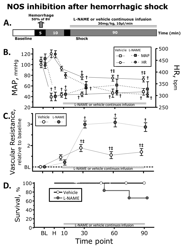
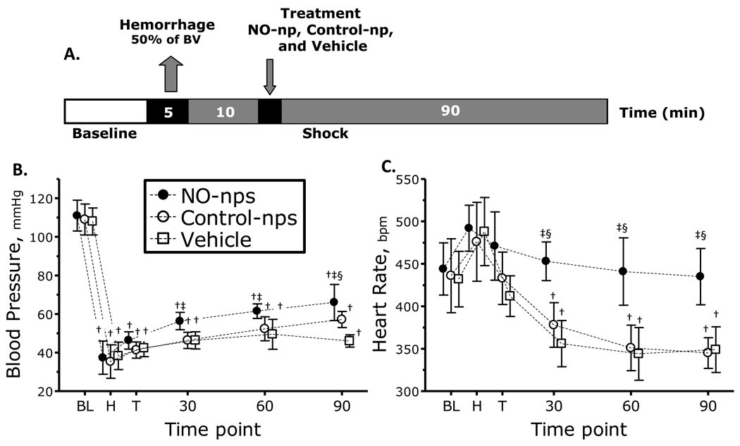
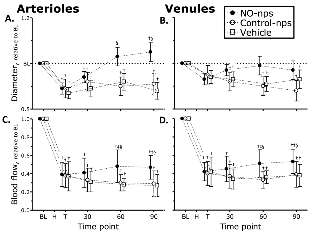
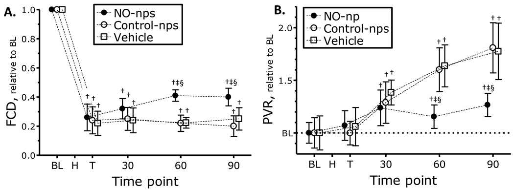
This study investigated the systemic and microvascular hemodynamic changes related to increased nitric oxide (NO) availability following significant hemorrhage, made available by administration of NO releasing nanoparticles (NO-nps). Hemodynamic responses to hemorrhagic shock were studied in the hamster window chamber. Acute hemorrhage was induced by arterial controlled bleeding of 50% of blood volume, and the resulting hemodynamic parameters were followed over 90 min. Exogenous NO was administered in the form of NO-nps (5mg/kg suspended in 50 μl saline) 10 min following induced hemorrhage. Control groups received equal dose of NO free nanoparticles (Control-nps) and Vehicle solution. Animals treated with NO-nps partially maintained systemic and microvascular function during hypovolemic shock compared to animals treated with Control-nps or the Vehicle (50 μl saline). The continuous NO released by the NO-nps reverted arteriolar vasoconstriction, partially recovered both functional capillary density and microvascular blood flows. Additionally, NO supplementation post hemorrhage prevented cardiac decompensation, and thereby maintained and stabilized the heart rate. Paradoxically, the peripheral vasodilation induced by the NO-nps did not decrease blood pressure, and combined with NO's effects on vascular resistance, NO-nps promoted intravascular pressure redistribution and blood flow, avoiding tissue ischemia. Therefore, by increasing NO availability with NO-nps during hypovolemic shock, it is possible that cardiac stability and microvascular perfusion can be preserved, ultimately increasing survivability and local tissue viability, and reducing hemorrhagic shock sequelae. The relevance, stability, and efficacy of exogenous NO therapy in the form of NO-nps will potentially facilitate the intended use in battlefield and trauma situations.


Conflict of interest statement

Conflict of interest statement

No conflicts of interest to declare.

Figures

Figure 1
Figure 1
Nitric oxide (NO) synthase (NOS) inhibition during hemorrhagic shock. A. Shock protocol. Hemorrhagic shock was induced withdrawal of 50% of the estimated blood volume (7% body weight) and NOS inhibition with L-NAME was started 10 min after the end of the hemorrhage. B. NOS inhibition effects on mean arterial pressure (MAP) and heart rate (HR) during the hemorrhagic shock. C. NOS inhibition effects on vascular resistance during the hemorrhagic shock. D. NOS inhibition effects on survivability during the hemorrhagic shock. †, P<0.05 compared to baseline; ‡, P<0.05 compared to Vehicle
Figure 2
Figure 2
A. Diagram hemorrhagic shock protocol. Hemorrhagic shock was induced withdrawal of 50% of the estimated blood volume (7% body weight). Treatments were administered 10 min after the end of the hemorrhage. B. and C. nitric oxide (NO) supplementation effects on mean arterial pressure (MAP) and heart rate (HR) during hemorrhagic shock. †, P<0.05 compared to baseline; ‡, P<0.05 compared to Vehicle; §, P<0.05 compared to Control-nps. Time points: Bl, baseline; H, after hemorrhage, T, after treatment, and 30, 60 and 90 min after treatment. MAP (mmHg, mean ± SD) for each animal group were as follows: Baseline: Vehicle, 108 ± 7, n = 6; Control-nps, 109 ± 8 n = 6; NO-nps, 111 ± 8, n = 6. n = number of animals. HR (bpm, mean ± SD) at baseline for each animal group was as follows: Vehicle, 432 ± 32; Control-nps, 436 ± 43; NO-nps, 444 ± 31.
Figure 3
Figure 3
Nitric oxide (NO) supplementation effects on changes in arteriolar and venular diameter (A. and B.) and blood flow (C. and D.) during hemorrhagic shock. Broken line represents baseline level. Time points: Bl, baseline; H, after hemorrhage, T, after treatment, and 30, 60 and 90 min after treatment. †, P<0.05 compared to baseline; ‡, P<0.05 compared to Vehicle; §, P<0.05 compared to Control-nps. Diameters (µm, mean ± SD) in Figures 3A (arteriolar) and 3B (venular) for each animal group were as follows: Baseline: Vehicle (arterioles (A): 62.7 ± 8.2, n = 26; venules (V): 64.5 ± 6.8, n = 24); Control-nps (A: 60.5 ± 6.8, n = 24; V: 65.7 ± 8.7, n = 27); NO-nps (A: 62.0 ± 7.4, n = 25, V: 64.5 ± 8.2, n = 26). n = number of vessels studied. RBC velocities (mm/s, mean ± SD for each animal group were as follows: Baseline: Vehicle (A: 4.3 ± 1.0, V: 2.3 ± 0.9); Control-nps (A: 4.5 ± 0.8; V: 2.4 ± 1.0); NO-nps (A: 4.3 ± 1.0; V: 2.6 ± 0.7). Calculated flows (nl/s, mean ± SD) in Figures 3C (arteriolar) and 3D (venular) for each animal group were as follows: Baseline: Vehicle (A: 11.7 ± 3.4; V: 6.9 ± 2.2); Control-nps (A: 12.1 ± 3.2; V: 7.1 ± 2.3); NO-nps (A: 12.0 ± 2.8; V: 6.8 ± 2.3).
Figure 4
Figure 4
A. Nitric oxide (NO) supplementation effects on changes in functional capillary density (FCD) and B. estimated peripheral vascular resistance (PVR) during hemorrhagic shock. Broken line represents baseline level. Estimation of PVR was made using Hagen-Poiseuille equation, with MAP and microvascular blood flow. Time points: Bl, baseline; H, after hemorrhage, T, after treatment, and 30, 60 and 90 min after treatment. †, P<0.05 compared to baseline; ‡, P<0.05 compared to Vehicle; §, P<0.05 compared to Control-nps. FCD (capillaries per cm, mean ± SD) for each animal group were as follows: Baseline: Vehicle, 111 ± 9; Control-nps, 112 ± 11; NO-nps, 109 ± 8.

References

    1. Bellamy RF. The causes of death in conventional land warfare: implications for combat casualty care research. Mil Med. 1984;149:55–62. - PubMed
    1. Pope AM Institute of Medicine (U.S.) Fluid resuscitation : state of the science for treating combat casualties and civilian injuries. Washington, D.C.: National Academy Press; 1999. - PubMed
    1. Bacter CR, Canizaro PC, Carrico CJ, Shires GT. Fluid resuscitation of hemorrhagic shock. Postgrad Med. 1970;48:95–99. - PubMed
    1. Burris D, Rhee P, Kaufmann C, Pikoulis E, Austin B, Eror A, et al. Controlled resuscitation for uncontrolled hemorrhagic shock. J Trauma. 1999;46:216–223. - PubMed
    1. Dubick MA, Atkins JL. Small-volume fluid resuscitation for the far-forward combat environment: current concepts. J Trauma. 2003;54:S43–S45. - PubMed

Publication types

MeSH terms