. 2011 Apr 15;26(8):3432-7.
doi: 10.1016/j.bios.2011.01.019. Epub 2011 Jan 22.

Label-free multiplexed virus detection using spectral reflectance imaging

Affiliations

Label-free multiplexed virus detection using spectral reflectance imaging

Carlos A Lopez et al. Biosens Bioelectron. .

Abstract

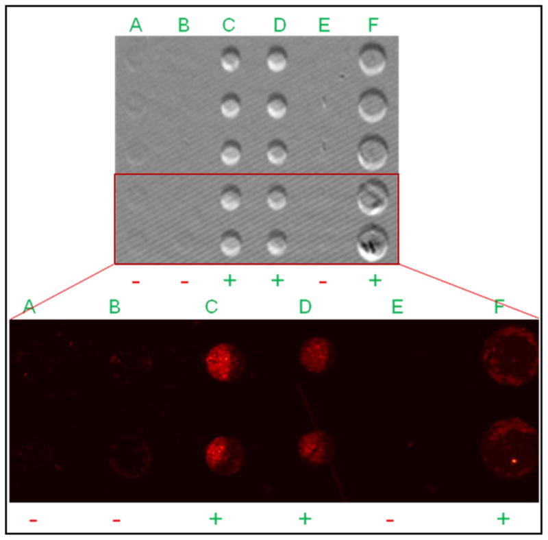
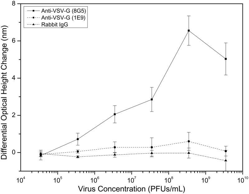
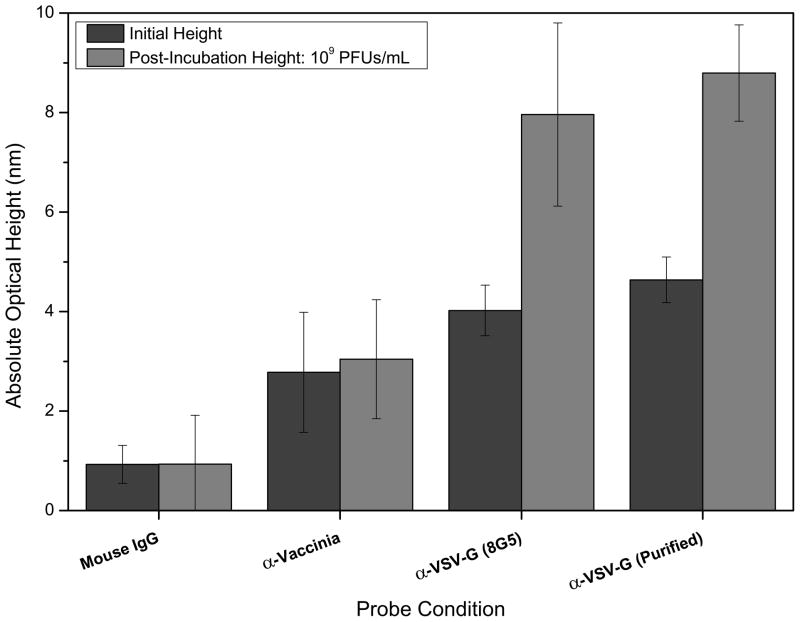
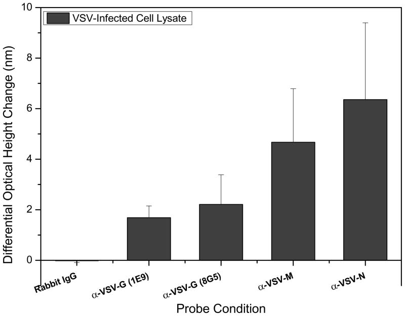
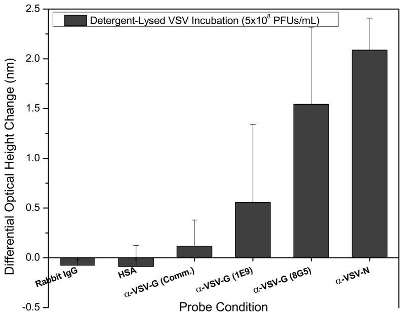
We demonstrate detection of whole viruses and viral proteins with a new label-free platform based on spectral reflectance imaging. The Interferometric Reflectance Imaging Sensor (IRIS) has been shown to be capable of sensitive protein and DNA detection in a real time and high-throughput format. Vesicular stomatitis virus (VSV) was used as the target for detection as it is well-characterized for protein composition and can be modified to express viral coat proteins from other dangerous, highly pathogenic agents for surrogate detection while remaining a biosafety level 2 agent. We demonstrate specific detection of intact VSV virions achieved with surface-immobilized antibodies acting as capture probes which is confirmed using fluorescence imaging. The limit of detection is confirmed down to 3.5 × 10(5)plaque-forming units/mL (PFUs/mL). To increase specificity in a clinical scenario, both the external glycoprotein and internal viral proteins were simultaneously detected with the same antibody arrays with detergent-disrupted purified VSV and infected cell lysate solutions. Our results show sensitive and specific virus detection with a simple surface chemistry and minimal sample preparation on a quantitative label-free interferometric platform.


Figures

Figure 1
Figure 1
(Top) The IRIS system showing the CCD camera, x-y-z sample stage, and light source and optical components required for illumination of the sensing area (antibody array). (Bottom) Schematic of the layered Si-SiO2 substrate spotted with a representative antibody array. Each antibody ensemble is spotted in replicate with specificity for a different epitope targeting different viral coat proteins (whole virus detection) or internal proteins (lysed virus detection). Negative control antibodies depend on the assay and can be non-specific and virus-specific.
Figure 2
Figure 2
Qualitative confirmation of label-free detection of whole WT VSV. Fluorescently-labeled VSV was incubated with a surface that had immobilized: A) mouse IgG, B) anti-Vaccinia, C & D) anti-VSV (clone 8G5), E) blank area control, F) anti-VSV (clone 1E9). A magnified fluorescence image shown below the label-free image demonstrates a correlation to the IRIS data for the exact same area, with positive and negative signs indicating significant and insignificant binding respectively. Greater fluorescence was observed for the expected probes (C, D, & F) while minimal fluorescence, indicating some non-specific binding, was observed for the other two probes (A, B) confirming the measured label-free signal.
Figure 3
Figure 3
Limit of detection analysis of the IRIS platform for whole VSV virion detection. Increasing concentrations of WT VSV solutions demonstrated corresponding changes in the spot heights of the two monoclonal, viral glycoprotein-specific antibody probes. Greater binding was observed for monoclonal 8G5 over the 1E9 clone indicating that antibody (probe) affinity is an important factor in increasing the assay LOD. Minimal non-specific binding was observed for the control rabbit IgG spots for all virus concentrations. The binding assay protocol used here results in a LOD close to 105 PFUs/mL.
Figure 4
Figure 4
Detection of whole VSV in a complex sample (DMEM containing 7% FBS). At a target concentration of 109 PFUs/mL, significant spot height changes were observed for the glycoprotein specific antibody probes, with minimal non-specific binding to the Vaccinia antibody and mouse IgG control probes. Two preparations of the anti-VSV-G 8G5 monoclonal were tested to determine if purification could improve probe functionality.
Figure 5
Figure 5
IRIS detection of external and internal viral proteins in VSV-infected cell lysate solutions. Antibodies specific for the internal matrix (M) and nucleocapsid (N) proteins, two different monoclonal antibodies for the coat glycoprotein (G), and a non-specific rabbit IgG control solution were spotted. Significant detection was achieved for all virus-specific probes with the greatest changes observed for the internal proteins. A negligible height change was observed for the mean of the control spots indicating that there was no binding to the control rabbit IgG spots.
Figure 6
Figure 6
IRIS protein detection of detergent-disrupted VSV virus solutions. Two different monoclonal antibodies for the coat glycoprotein (G), a commercially-available polyclonal for VSV-G, and an antibody targeted at the internal nucleocapsid (N) protein were used as specific probes. Non-specific rabbit IgG and human serum albumin (HSA) solutions were spotted as control probes. Viral lysis and subsequent protein release was achieved with a solution containing 0.5% NP-40. Significant detection of G and N proteins without non-specific binding to the control probes was achieved at a VSV concentration of 5×10⁸ PFUs/mL. The commercially available antibody specific for VSV-G demonstrated minimal binding of approximately 0.12 nm.

References

    1. Stamboulian D, Bonvehi PE, Nacinovich FM, Cox N. Infect Dis Clin North Am. 2000;14 (1):141–166. - PubMed
    1. Amano Y, Cheng Q. Anal Bioanal Chem. 2005;381 (1):156–164. - PubMed
    1. Charlton B, Crossley B, Hietala S. Comp Immunol Microbiol Infect Dis. 2009;32 (4):341–350. - PubMed
    1. Baner J, Gyarmati P, Yacoub A, Hakhverdyan M, Stenberg J, Ericsson O, Nilsson M, Landegren U, Belak S. J Virol Methods. 2007;143 (2):200–206. - PubMed
    1. Baxi MK, Baxi S, Clavijo A, Burton KM, Deregt D. Vet J. 2006;172 (3):473–481. - PubMed

Publication types

MeSH terms

LinkOut - more resources