. 2011 May;152(5):2011-9.
doi: 10.1210/en.2010-0871. Epub 2011 Feb 22.

Spatially selective, testosterone-independent remodeling of dendrites in gonadotropin-releasing hormone (GnRH) neurons prepubertally in male rats

Affiliations

Spatially selective, testosterone-independent remodeling of dendrites in gonadotropin-releasing hormone (GnRH) neurons prepubertally in male rats

Natividad Ybarra et al. Endocrinology. 2011 May.

Abstract

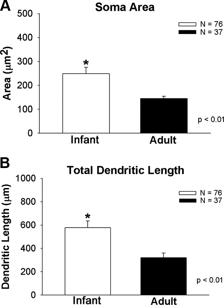
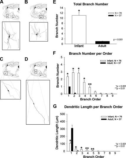
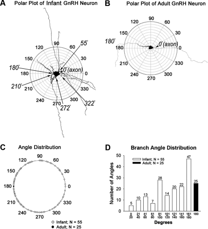
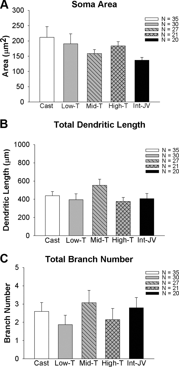
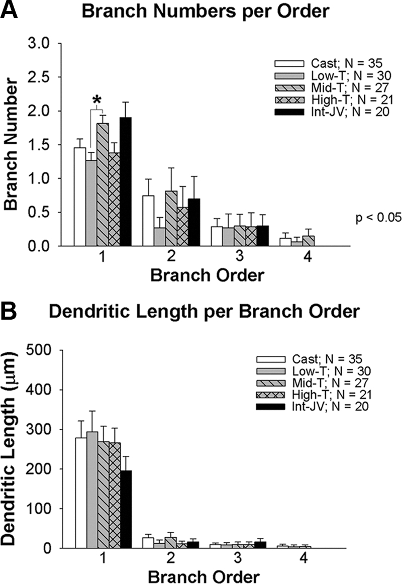
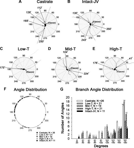
Adult GnRH neurons exhibit a stereotypic morphology with a small soma, single axon, and single dendrite arising from the soma with little branching. The adult morphology of GnRH neurons in mice reflects an anatomical consolidation of dendrites over postnatal development. We examined this issue in rat GnRH neurons with biocytin filling in live hypothalamic slices from infant males, as adult littermates and in gonad-intact males, castrated males, and in males with one of three levels of testosterone (T) treatment. Somatic area and total dendritic length were significantly greater in infant males than in adults. Moreover, total numbers of dendrite branches were greater in infant males as compared with adults. The number of higher order branches and the lengths of higher order branches were also greater in infant males than in adults. Most interestingly, in adults a single dendrite arose from the somata, consistently at 180° from the axon. In contrast, prepubertal animals had an average of 2.2 ± 0.2 primary dendrites arising from somata (range, one to seven primary dendrites). Angles relative to the axon at which dendrites in prepubertal males emanated from GnRH somata were highly variable. Castration at 25 d of age and castration at 25 d of age with one of three levels of T treatment did not influence morphological parameters when GnRH neurons were examined between 40 d and 48 d of age. Thus, a spatially selective remodeling of primary dendrites and consolidation of distal GnRH dendritic arbors occurs during postnatal development and is largely independent of T.


Figures

Fig. 1.
Fig. 1.
A, Differences in somatic area between GnRH neurons derived from infant and adult males. B, Differences in total dendritic length between GnRH neurons derived from infant and adult males. Total dendritic length is the additive sum of all primary dendrites and associated branches for any given neuron. Asterisks indicate a significant difference between groups.
Fig. 2.
Fig. 2.
Morphological differences between dendrites of GnRH neurons derived from infant and adult males. Top portion of each panel (A–D) indicates the anatomical location of representative biocytin-filled GnRH neurons shown in the bottom panel. Infant GnRH neurons are shown in A and B. Adult GnRH neurons are shown in C and D. Scale bars, 100 μm. E, Indicates that the total numbers of branches is higher in infant males than adults. There is also more, higher order branching in infant GnRH neurons than adult GnRH neurons (F). Dendritic length per branch order is shown in G. Branch order and length comparisons were only performed between infant and adult groups where branches existed in both groups. Asterisks indicate significant differences as noted by P values.
Fig. 3.
Fig. 3.
Polar plot analysis of an infant (A) and an adult GnRH neuron (B). For polar plots, the axon of each GnRH neuron was aligned to 0°. The angles at which dendrites emerged from the soma were then determined and plotted on a single diagram (C) for infant (shown in white circles) and for adult (shown in black circle) males. D, Distribution of dendritic angles for primary dendrites. Only neurons in which clear axons emerged from somata were included in this analysis.
Fig. 4.
Fig. 4.
Somatic area (A), total dendritic length (B), and total branch number (C) in GnRH neurons derived from juvenile males that were castrated at 25 d of age and were either implanted with empty implants (castrates) or implants containing one of three levels of T. A group of intact males was also studied. T levels are shown in Table 1.
Fig. 5.
Fig. 5.
Total numbers of branches per order (A) and dendritic length per branch order (B) of dendrites in GnRH neurons from juvenile males that were castrated at 25 d of age and were either implanted with empty implants (Cast) or implants containing one of three levels of T (Low-T, Mid-T, High-T). A group of intact juvenile males (Int-JV) was also studied. T levels are shown in Table 1. Asterisks indicate a significant difference between groups.
Fig. 6.
Fig. 6.
Polar plot analysis of GnRH neurons derived from juvenile males that were castrated at 25 d of age were implanted with empty implants (A), left intact and not treated (B), or castrated and treated with implants containing one of three levels of T (C–E). For polar plots, the axon of each GnRH neuron was aligned to 0°. The angle at which dendrites emerged from the soma was then determined and plotted on a single diagram (F). G, Shows the distribution of dendritic angles. Only neurons in which clear axons emerged from somata were included in this analysis. JV, Juvenile.

References

    1. Urbanski HF, Ojeda SR. 1987. Activation of luteinizing hormone-releasing hormone release advances the onset of female puberty. Neuroendocrinology 46:273–276 - PubMed
    1. Harris GC, Levine JE. 2003. Pubertal acceleration of pulsatile gonadotropin-releasing hormone release in male rats as revealed by microdialysis. Endocrinology 144:163–171 - PubMed
    1. Merchenthaler I, Görcs T, Sétáló G, Petrusz P, Flerkó B. 1984. Gonadotropin-releasing hormone (GnRH) neurons and pathways in the rat brain. Cell Tissue Res 237:15–29 - PubMed
    1. Silverman A-J, Livne I, Witkin JW. 1994. The gonadotropin releasing-hormone (GnRH) neuronal systems: immunocytochemistry and in situ hybridization. In: Knobil E, Neill JD. eds. The physiology of reproduction. New York: Raven Press; 1683–1710
    1. Jennes L. 1989. Prenatal development of the gonadotropin-releasing hormone-containing systems in rat brain. Brain Res 482:97–108 - PubMed

Publication types

MeSH terms