. 2011 Apr 22;286(16):13966-76.
doi: 10.1074/jbc.M110.186338. Epub 2011 Feb 22.

Sequence-independent control of peptide conformation in liposomal vaccines for targeting protein misfolding diseases

Affiliations

Sequence-independent control of peptide conformation in liposomal vaccines for targeting protein misfolding diseases

David T Hickman et al. J Biol Chem. .

Abstract

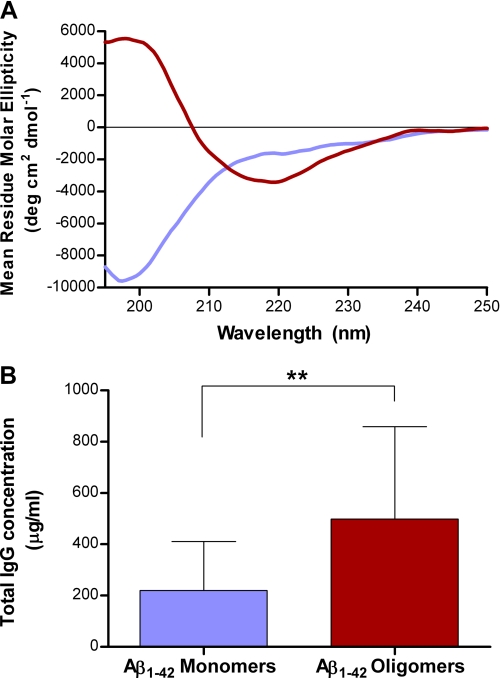
Synthetic peptide immunogens that mimic the conformation of a target epitope of pathological relevance offer the possibility to precisely control the immune response specificity. Here, we performed conformational analyses using a panel of peptides in order to investigate the key parameters controlling their conformation upon integration into liposomal bilayers. These revealed that the peptide lipidation pattern, the lipid anchor chain length, and the liposome surface charge all significantly alter peptide conformation. Peptide aggregation could also be modulated post-liposome assembly by the addition of distinct small molecule β-sheet breakers. Immunization of both mice and monkeys with a model liposomal vaccine containing β-sheet aggregated lipopeptide (Palm1-15) induced polyclonal IgG antibodies that specifically recognized β-sheet multimers over monomer or non-pathological native protein. The rational design of liposome-bound peptide immunogens with defined conformation opens up the possibility to generate vaccines against a range of protein misfolding diseases, such as Alzheimer disease.


Figures

FIGURE 1.
FIGURE 1.
Conformational analyses and metal binding properties of liposomal Palm1–15 peptide ACI-24 composed of Palm1–15 (121 μm), DMPC (10.9 mm), DMPG (1.21 mm), cholesterol (8.51 mm), and MPLA (77 μm). A, fluorescence emission of liposomal formulation of Palm1–15 peptide (ACI-24) (□) and upon the addition of ThT to ACI-24 (■), ACI-24-Empty (▴), and Acetyl1–15 peptide (×). Samples were analyzed in PBS at a peptide concentration of 15 μm with excitation at 440 nm. B, ThT binding isotherm for liposomal peptide Palm1–15 (ACI-24). C, CD spectra of lipidated peptide Palm1–15 embedded in liposomes (solid line) and of acetylated “native” peptide Acetyl1–15 (dotted line) in PBS. Spectra were recorded after 7-fold dilution for liposomal Palm1–15 in PBS with subtraction of a spectrum of the corresponding empty liposomes lacking peptide. D, fluorescence emission of ThT in the presence of liposomal vaccine ACI-24 upon the addition of different metal ions. ThT was added to ACI-24 alone or to ACI-24 (final concentration 15 μm) preincubated with different metals (final concentration 20 μm) prior to the addition of metal chelator diethylenetriamine pentaacetic acid (DPTA) (final concentration 200 μm). As a control, diethylenetriamine pentaacetic acid was added to liposomal Palm1–15 in the absence of Cu(II), resulting in no change to the ThT emission. Data are expressed as average ± S.D. (error bars) (n = 3). RFU, relative fluorescence units.
FIGURE 2.
FIGURE 2.
Conformational analyses of various N- and C-terminal dipalmitoylated peptides of different primary sequence. A, CD spectra of liposomal peptides Palm15–1 (solid line), scPalm15 (dashed line), Palm1–9 (×), and Palm1–5 (dotted line). B, ThT fluorescence emission upon the addition to liposomes embedded with different sequences of N- and C-dipalmitoylated peptides (filled bars) and corresponding “native” acetylated peptides (open bars) in the absence of liposomes. Results are given as average ± S.D. (error bars) (n = 3). C, CD spectra of liposomal peptides Palm1–15(D7K) (solid line), Palm1–15(E3A,D7K) (dashed line), Palm1–15(E3K,D7K) (×), and Palm1–15(E3K,D7K,E11K) (dotted line). RFU, relative fluorescence units.
FIGURE 3.
FIGURE 3.
Effect of liposome surface charge upon conformation of N- and C-terminal dipalmitoylated peptides integrated into liposomal bilayers. A, effect of liposome surface charge upon conformation of Palm1–15 peptide (■, liposome formulations A–F; see supplemental Table S3), and cationic mutant Palm1–15(E3K,D7K,E11K) (△, liposome formulations G–L) measured by ThT fluorescence. Liposomes are composed of peptide, phospholipids (different proportions of DMPC, DMPG, and/or DMTAP), cholesterol, and MPLA in a molar ratio of 0.1:10:7:0.06, respectively. Empty liposomes lacking peptide (●, liposome formulations M–O) were used as negative controls. ζ potential values are given as average ± S.D. (n = 3). B, 13C-13C double quantum MAS-NMR spectra of Palm1–15(ASG) peptide uniformly (13C/15N) labeled at positions Ala-2, Ser-8, and Gly-9 and incorporated into anionic liposomes (DMPC/DMPG/cholesterol/MPLA (molar ratio 9:1:7:0.06)) (dark blue line) or cationic liposomes (DMTAP/cholesterol/MPLA (molar ratio 10:7:0.06)) (dark red line). Conformation-dependent chemical shift ranges (36) compatible with α-helix (blue), random coil (red), and β-sheet (green) backbone structure are annotated. RFU, relative fluorescence units. CA and CB indicate the α- and β-Z carbon atoms of the corresponding amino acids.
FIGURE 4.
FIGURE 4.
Secondary and quaternary structural analysis of liposomal lipopeptides. A, ThT fluorescence upon the addition to liposomal peptide constructs Palm1–15(4C) (○), Palm1–15 (■), Palm1–15(2C) (▴), Palm1–15(1N1C) (△), and Palm1–15(1C) (□). B, CD spectra of liposomal peptide constructs Palm(4C) (solid line), Palm1–15(2C) (×), Palm1–15(1N1C) (dashed line), and Palm1–15(1C) (dotted line). C, ThT fluorescence upon the addition to liposomal peptide constructs Dodecyl1–15 (▴), Octyl1–15 (■), and Butyl1–15 (●). D, CD spectra of liposomal peptide constructs Dodecyl1–15 (dashed line), Octyl1–15 (solid line), and Butyl1–15 (dotted line). RFU, relative fluorescence units.
FIGURE 5.
FIGURE 5.
Conformation-specific binding of anti-ACI-24 polyclonal antibodies to Aβ(1–42). A, CD spectra of SEC purified oligomer (red) and monomer fractions (blue) of Aβ(1–42). B, recognition of distinct Aβ(1–42) conformations by polyclonal plasma taken from WT mice after immunization with liposomal vaccine ACI-24. Monomeric (random coil) and oligomeric (β-sheet) Aβ(1–42) species were coated onto an ELISA plate, and binding of plasma was quantified relative to control IgG antibody 6E10. Data are expressed in mean ± S.D. (error bars) (n = 10). **, p < 0.01 by non-parametric Wilcoxon matched pairs ranks.
FIGURE 6.
FIGURE 6.
Staining of pathological misfolded β-sheet plaques in human AD brain. Shown is staining of human AD brain (A) and healthy human brain (B), with sera (diluted 1:2000) taken from a monkey immunized with ACI-24. Shown is staining of human AD brain with positive control monoclonal antibody (Dako mouse mAb) (C) or with sera from a monkey immunized with PBS (D). Brain slices are magnified 40-fold.

References

    1. Misumi S., Nakayama D., Kusaba M., Iiboshi T., Mukai R., Tachibana K., Nakasone T., Umeda M., Shibata H., Endo M., Takamune N., Shoji S. (2006) J. Immunol. 176, 463–471 - PubMed
    1. Villard V., Agak G. W., Frank G., Jafarshad A., Servis C., Nébié I., Sirima S. B., Felger I., Arevalo-Herrera M., Herrera S., Heitz F., Bäcker V., Druilhe P., Kajava A. V., Corradin G. (2007) PLoS ONE 2, e645–647 - PMC - PubMed
    1. Su H. P., Golden J. W., Gittis A. G., Hooper J. W., Garboczi D. N. (2007) Virology 368, 331–341 - PMC - PubMed
    1. Pimentel T. A., Yan Z., Jeffers S. A., Holmes K. V., Hodges R. S., Burkhard P. (2009) Chem. Biol. Drug Des. 73, 53–61 - PMC - PubMed
    1. Gao J., Sidhu S. S., Wells J. A. (2009) Proc. Natl. Acad. Sci. U.S.A. 106, 3071–3076 - PMC - PubMed