

Computer-aided detection of breast masses depicted on full-field digital mammograms: a performance assessment
- PMID: 21343322
- PMCID: PMC3120913
- DOI: 10.1259/bjr/51461617
Computer-aided detection of breast masses depicted on full-field digital mammograms: a performance assessment
Abstract
Objectives: To investigate the feasibility of converting a computer-aided detection (CAD) scheme for digitised screen-film mammograms to full-field digital mammograms (FFDMs) and assessing CAD performance on a large database.
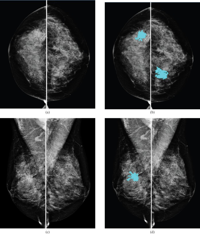
Methods: The database included 6478 FFDM images acquired on 1120 females, with 525 cancer cases and 595 negative cases. The database was divided into five case groups: (1) cancer detected during screening, (2) interval cancers, (3) "high-risk" recommended for surgical excision, (4) recalled but negative and (5) negative (not recalled). A previously developed CAD scheme for masses depicted on digitised images was converted and re-optimised for FFDM images while keeping the same image-processing structure. CAD performance was analysed on the entire database.
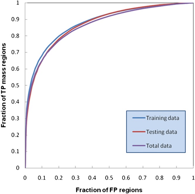
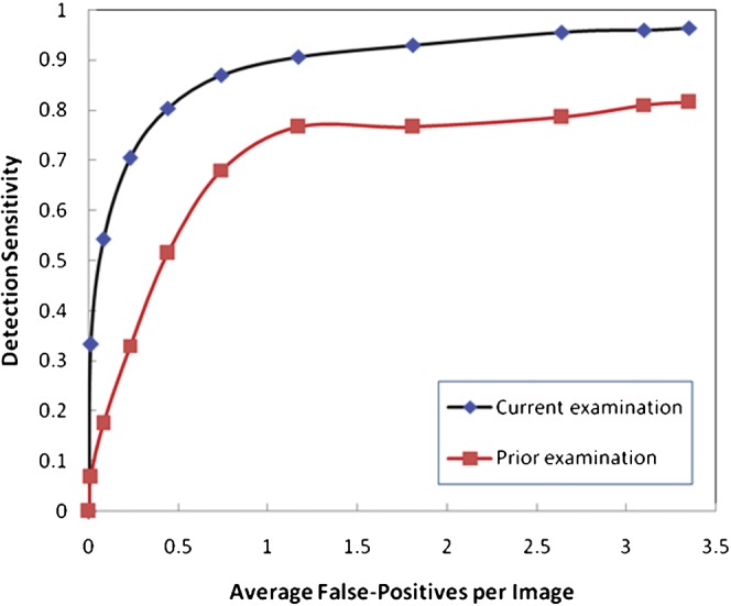
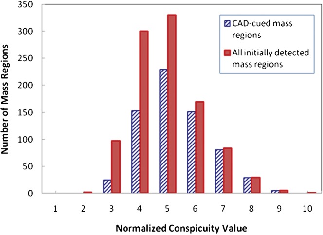
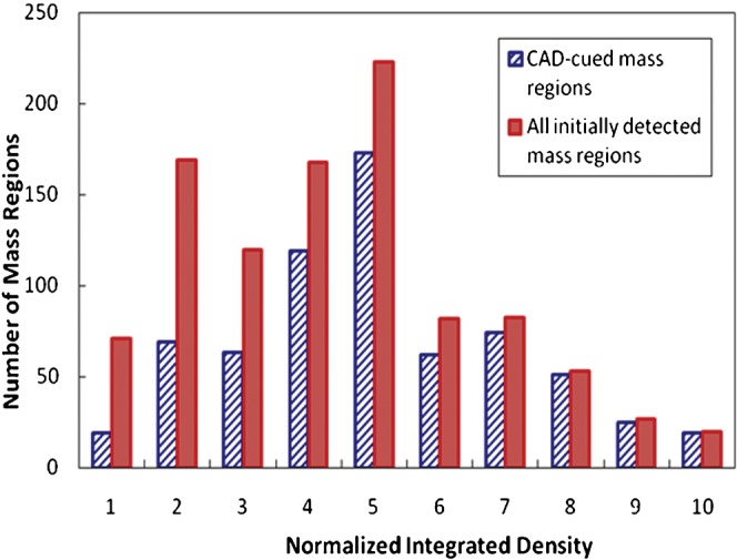
Results: The case-based sensitivity was 75.6% (397/525) for the current mammograms and 40.8% (42/103) for the prior mammograms deemed negative during clinical interpretation but "visible" during retrospective review. The region-based sensitivity was 58.1% (618/1064) for the current mammograms and 28.4% (57/201) for the prior mammograms. The CAD scheme marked 55.7% (221/397) and 35.7% (15/42) of the masses on both views of the current and the prior examinations, respectively. The overall CAD-cued false-positive rate was 0.32 per image, ranging from 0.29 to 0.51 for the five case groups.
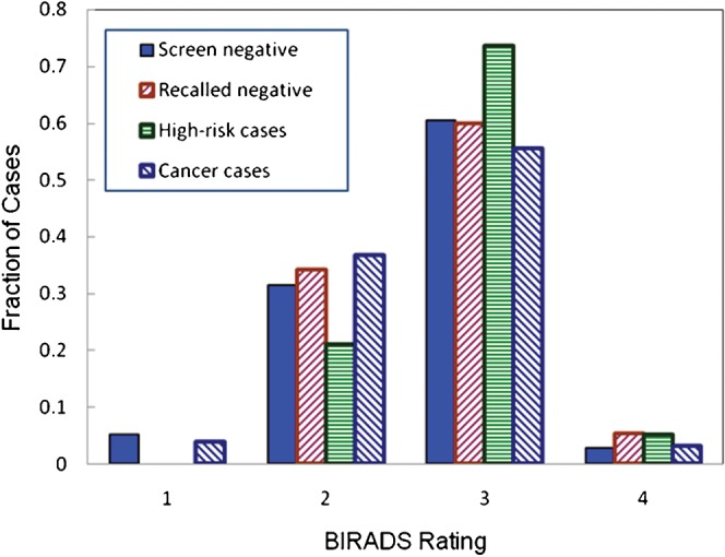
Conclusion: This study indicated that (1) digitised image-based CAD can be converted for FFDMs while performing at a comparable, or better, level; (2) CAD detects a substantial fraction of cancers depicted on prior examinations, albeit most having been marked only on one view; and (3) CAD tends to mark more false-positive results on "difficult" negative cases that are more visually difficult for radiologists to interpret.
Figures
References
-
- Freer TM, Ulissey MJ. Screening mammography with computer-aided detection: prospective study of 12,860 patients in a community breast center. Radiology 2001;220:781–6 - PubMed
-
- Gur D, Sumkin JH, Rockette HE, Ganott M, Hakim C, Hardesty L, et al. Changes in breast cancer detection and mammography recall rates after the introduction of a computer-aided detection system. J Natl Cancer Inst 2004;96:185–90 - PubMed
-
- Khoo LA, Taylor P, Given-Wilson RM. Computer-aided detection in the United Kingdom National Breast Screening Programme: prospective study. Radiology 2005;237:444–9 - PubMed
-
- Ko JM, Nicholas MJ, Mendel JB, Slanetz PJ. Prospective assessment of computer-aided detection in interpretation of screening mammograms. AJR Am J Roentgenol 2006;187:1483–91 - PubMed
Publication types
MeSH terms
Grants and funding
LinkOut - more resources
Full Text Sources
Medical
Miscellaneous
