. 2011 May;31(9):1921-33.
doi: 10.1128/MCB.01130-10. Epub 2011 Feb 22.

Pathways for Holliday junction processing during homologous recombination in Saccharomyces cerevisiae

Affiliations

Pathways for Holliday junction processing during homologous recombination in Saccharomyces cerevisiae

Thomas M Ashton et al. Mol Cell Biol. 2011 May.

Abstract

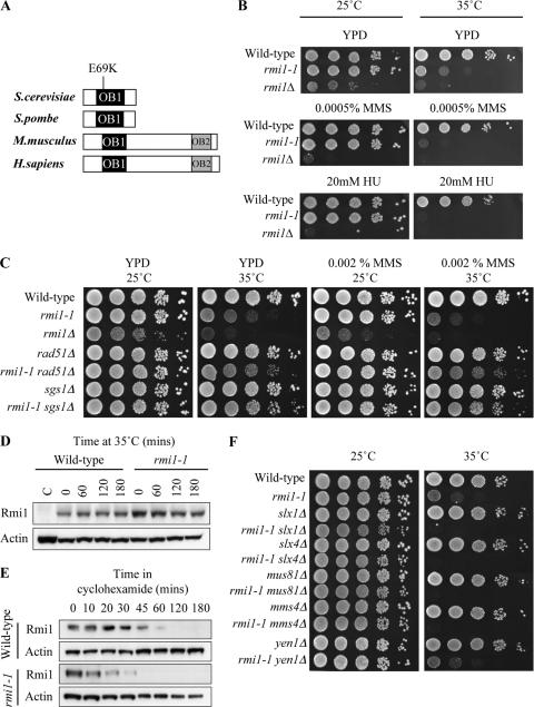
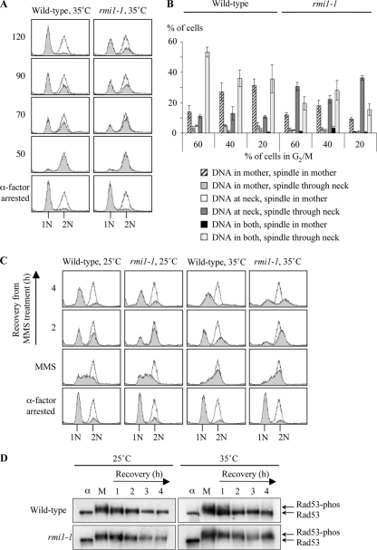
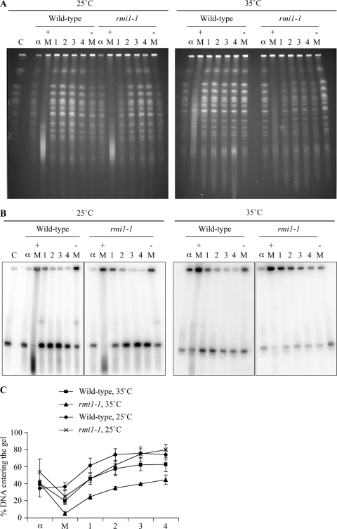
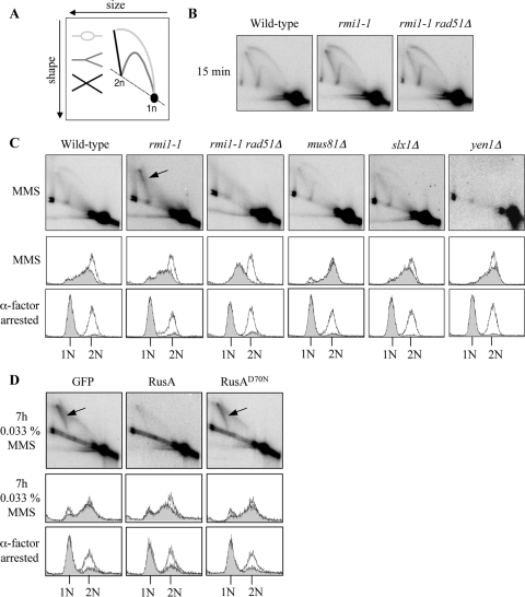
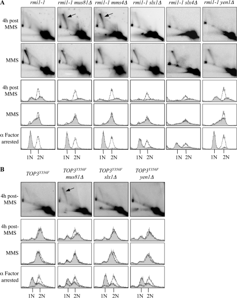
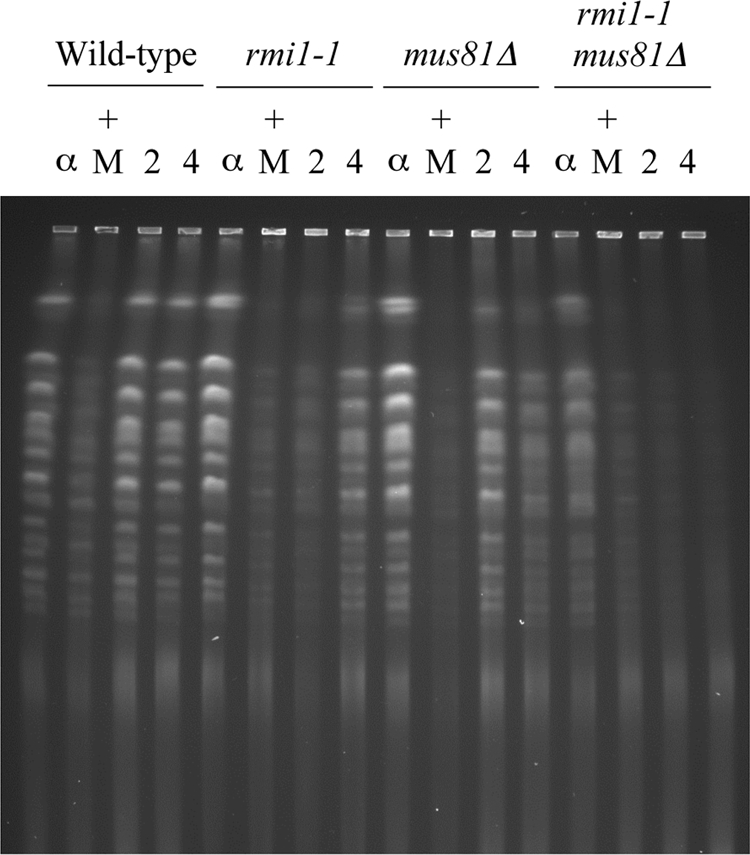
The Saccharomyces cerevisiae Rmi1 protein is a component of the highly conserved Sgs1-Top3-Rmi1 complex. Deletion of SGS1, TOP3, or RMI1 is synthetically lethal when combined with the loss of the Mus81-Mms4 or Slx1-Slx4 endonucleases, which have been implicated in Holliday junction (HJ) resolution. To investigate the causes of this synthetic lethality, we isolated a temperature-sensitive mutant of the RMI1 strain, referred to as the rmi1-1 mutant. At the restrictive temperature, this mutant phenocopies an rmi1Δ strain but behaves like the wild type at the permissive temperature. Following a transient exposure to methyl methanesulfonate, rmi1-1 mutants accumulate unprocessed homologous recombination repair (HRR) intermediates. These intermediates are slowly resolved at the restrictive temperature, revealing a redundant resolution activity when Rmi1 is impaired. This resolution depends on Mus81-Mms4 but not on either Slx1-Slx4 or another HJ resolvase, Yen1. Similar results were also observed when Top3 function was impaired. We propose that the Sgs1-Top3-Rmi1 complex constitutes the main pathway for the processing of HJ-containing HRR intermediates but that Mus81-Mms4 can also resolve these intermediates.


Figures

Fig. 1.
Fig. 1.
rmi1-1 encodes a temperature-sensitive allele of RMI1. (A) RMI1 homologs aligned by the OB-fold domain OB1. The position of the E69K point mutation in the rmi1-1 allele is indicated. (B) rmi1-1 cells exhibit normal growth at 25°C but growth inhibition at 35°C. Growth of wild-type (BY4741), rmi1-1, and rmi1Δ strains was compared on plates in the absence of DNA-damaging agents (YPD) and on plates containing either 0.0005% MMS or 20 mM HU. Tenfold serial dilutions of each strain were spotted onto plates, and plates were incubated at 25°C for 3 days or at 35°C for 2 days. (C) Mutation of RAD51 or SGS1 suppresses rmi1-1 phenotypes. Growth of the indicated strains was compared on plates in the absence of DNA damaging agents (YPD) and on plates containing either 0.002% MMS or 20 mM HU. (D) Rmi1 is not degraded at 35°C in rmi1-1 cells. Protein extracts were prepared from asynchronous wild-type (C), RMI1-HA, and rmi1-1-HA strains grown at 25°C. The RMI1-HA and rmi1-1-HA strains were incubated at 37°C for 180 min. Levels of Rmi1 were analyzed by Western blotting. Actin was used as a loading control. (E) Degradation of Rmi1 is slightly faster in rmi1-1 cells when de novo protein synthesis is inhibited. RMI1-HA and rmi1-1-HA strains were arrested in G1 at 25°C and released into fresh medium. Once the cells had reached S phase, cycloheximide was added, and the cells were incubated at 35°C for 180 min. (F) Mutation of SLX1, SLX4, MUS81, and MMS4 causes synthetic lethality in rmi1-1 cells at 35°C.
Fig. 2.
Fig. 2.
rmi1-1 cells have an extended G2/M delay after completing a perturbed S phase but are proficient in DNA damage checkpoint activation. (A) rmi1-1 cells exhibit a mild G2/M delay. Wild-type and rmi1-1 strains were arrested in G1 (1N) at 35°C, washed, and resuspended in fresh medium. Cell cycle progression was followed by flow cytometry. α-Factor was added to the cultures at 50 min to prevent cells from entering a second S phase. The shaded peaks represent experimental data, and the unshaded peaks indicate a normal G2/M peak (2N). (B) Mitotic DNA segregation defects arise after traversing the first S phase in which functional Rmi1 is absent. Fifty cells were scored blindly in three independent experiments from the samples in which 60%, 40%, or 20% of the cells were in G2/M (60, 70, and 80 min in wild-type cells and 80, 90, and 120 min in rmi1-1 cells, respectively). The position of the nucleus was scored as follows: at the neck of the mother cell (at neck), elsewhere in the mother cell (in mother only), in the daughter cell only, or in both the mother and the daughter cells. The spindle was stained using an anti-α-tubulin antibody, and its position in each cell was also scored. (C) MMS exacerbates the G2/M delay in rmi1-1 cells. Wild-type and rmi1-1 cells were grown at 25°C, arrested in G1 at either 25°C or 35°C, and then released into medium containing 0.0167% MMS at the indicated temperature. After 1.5 h the MMS was washed off, and the cells were allowed to recover for 4 h in drug-free medium. α-Factor was added to prevent any cells traversing mitosis and entering a second round of DNA replication. (D) DNA damage checkpoint activation is proficient in rmi1-1 cells. Protein extracts were prepared from the cultures in Fig. 2C. Rad53 phosphorylation status was monitored by Western blotting. The positions of unphosphorylated Rad53 and slower-migrating phosphorylated forms of Rad53 are shown on the right. α, α-factor-arrested sample; M, sample taken after 1.5 h exposure to 0.0167% MMS. The numbers 1, 2, 3, and 4 refer to the number of hours after the MMS was removed from the cultures.
Fig. 3.
Fig. 3.
Branched DNA structures persist in rmi1-1 cells after a perturbed S phase. (A) Chromosome integrity is impaired following MMS treatment in rmi1-1 cells. Samples were taken from strains in Fig. 2C, and analyzed at the indicated times by pulsed-field gel electrophoresis (PFGE). C, yeast chromosomal DNA marker; α, α-factor-arrested sample; + M, sample taken after 1.5 h exposure to 0.0167% MMS; − M, sample taken after 50 min in the absence of MMS. 1, 2, 3 and 4 refer to the number of hours after the MMS was removed from the cultures. (B) Branched structures impair chromosome III electrophoretic mobility following MMS treatment in rmi1-1 cells. The DNA was transferred by Southern blotting and then hybridized with a probe that binds to ARS305 on chromosome III. (C) Quantification of chromosome III migration. The intensity of chromosome III in the wells versus the gel in panel B was quantified for each time point. The data are from three independent experiments. Error bars show standard errors.
Fig. 4.
Fig. 4.
Expression of the E. coli RusA HJ resolvase diminishes MMS-induced X-shaped DNA molecules in rmi1-1 cells. (A) DNA structures detectable using the 2D electrophoresis technique. Bubble (light gray), Y-shaped (dark gray), and X-shaped DNA molecules (black) are indicated. (B) Origin firing at ARS305 is unaffected in rmi1-1 cells. Wild-type, rmi1-1, and rmi1-1 rad51Δ strains were released from G1 arrest at 35°C into medium containing 0.0167% MMS. After 15 min, DNA replication bubbles, Y-shaped structures, and origin-associated, X-shaped DNA structures were detectable by 2D gel electrophoresis at the early-firing ARS305 origin on chromosome III. (C) RAD51-dependent, MMS-induced, X-shaped DNA molecules persist in rmi1-1 cells but not in strains lacking putative HJ resolvases. The indicated strains were released from G1 arrest at 35°C into medium containing 0.0167% MMS for 1.5 h. DNA replication intermediates were detected at ARS305 by 2D gel electrophoresis. RAD51-dependent X-shaped DNA molecules are indicated by the arrow. The lower panels indicate that wild-type, rmi1-1, mus81Δ, slx1Δ, and yen1Δ cells progress through S phase with similar kinetics, as measured by flow cytometry. (D) Overexpression of the RusA HJ resolvase diminishes MMS-induced X-shaped DNA molecules in rmi1-1 cells. rmi1-1-GFP, rmi1-1-RusA, and rmi1-1-RusAD70N strains were released from G1 arrest into medium containing 0.033% MMS. Protein expression was induced during the G1 arrest, and throughout the subsequent incubation, by the addition of 2% galactose. Cultures were released from G1 arrest at 35°C and were then incubated at 37°C after 1 h to promote robust HJ resolvase activity. Samples were taken for 2D gel electrophoresis after 7 h exposure to 0.033% MMS.
Fig. 5.
Fig. 5.
Inactivation of Top3 or Rmi1 in the absence of Mus81-Mms4 impairs the resolution of MMS-induced X-shaped DNA molecules. (A) Resolution of MMS-induced X-shaped DNA molecules is impaired in rmi1-1 cells lacking MUS81 or MMS4. The indicated strains were released from G1 arrest at 35°C into medium containing 0.0167% MMS for 1.5 h and then resuspended in drug-free medium as described for Fig. 2C. DNA replication intermediates arising at ARS305 were examined by 2D gel electrophoresis after a 1.5-h exposure to 0.0167% MMS and after 4 h of recovery following the removal of MMS. (B) Resolution of MMS-induced, X-shaped DNA molecules is impaired in mus81Δ cells overexpressing TOP3Y356F. The indicated strains were released from G1 arrest into medium containing 0.0167% MMS for 1.5 h and then resuspended in drug-free medium to allow recovery as described for Fig. 2C. DNA replication intermediates arising at ARS305 were examined by 2D gel electrophoresis after 4 h of recovery following the removal of MMS.
Fig. 6.
Fig. 6.
Deletion of MUS81 exacerbates defects in rmi1-1 mutants. The indicated strains were released from G1 arrest at 35°C into medium containing 0.0167% MMS for 1.5 h and then resuspended in drug-free medium as described for Fig. 2C. Samples were taken following α-factor arrest (α), after 1.5 h exposure to 0.0167% MMS (+ M), after 2 h of recovery from MMS treatment (2), and after 4 h of recovery (4). The samples were analyzed by PFGE.

References

    1. Amberg D., Burke D., Strathern J. 2005. Methods in yeast genetics. Cold Spring Harbor Laboratory Press, Cold Spring Harbor, NY
    1. Ashton T. M., Hickson I. D. 2010. Yeast as a model system to study RecQ helicase function. DNA Repair (Amsterdam) 9:303–314 - PubMed
    1. Bastin-Shanower S. A., Fricke W. M., Mullen J. R., Brill S. J. 2003. The mechanism of Mus81-Mms4 cleavage site selection distinguishes it from the homologous endonuclease Rad1-Rad10. Mol. Cell. Biol. 23:3487–3496 - PMC - PubMed
    1. Bellaoui M., et al. 2003. Elg1 forms an alternative RFC complex important for DNA replication and genome integrity. EMBO J. 22:4304–4313 - PMC - PubMed
    1. Bennett R. J., Keck J. L., Wang J. C. 1999. Binding specificity determines polarity of DNA unwinding by the Sgs1 protein of S. cerevisiae. J. Mol. Biol. 289:235–248 - PubMed

Publication types

MeSH terms

LinkOut - more resources