. 2011 May;31(9):1812-21.
doi: 10.1128/MCB.01149-10. Epub 2011 Feb 22.

RNA helicase p68 (DDX5) regulates tau exon 10 splicing by modulating a stem-loop structure at the 5' splice site

Affiliations

RNA helicase p68 (DDX5) regulates tau exon 10 splicing by modulating a stem-loop structure at the 5' splice site

Amar Kar et al. Mol Cell Biol. 2011 May.

Abstract

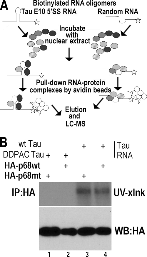
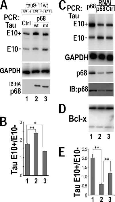
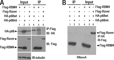
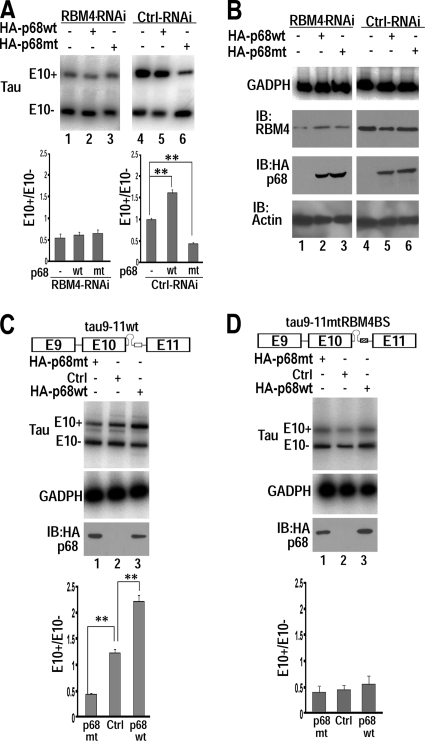
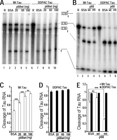
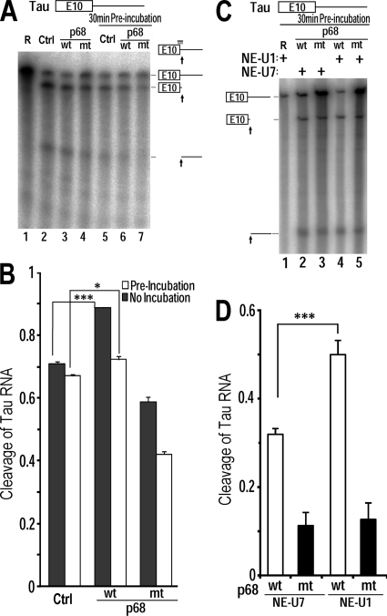
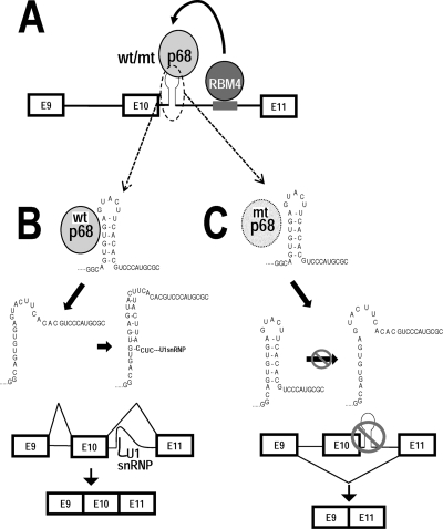
Regulation of tau exon 10 splicing plays an important role in tauopathy. One of the cis elements regulating tau alternative splicing is a stem-loop structure at the 5' splice site of tau exon 10. The RNA helicase(s) modulating this stem-loop structure was unknown. We searched for splicing regulators interacting with this stem-loop region using an RNA affinity pulldown-coupled mass spectrometry approach and identified DDX5/RNA helicase p68 as an activator of tau exon 10 splicing. The activity of p68 in stimulating tau exon 10 inclusion is dependent on RBM4, an intronic splicing activator. RNase H cleavage and U1 protection assays suggest that p68 promotes conformational change of the stem-loop structure, thereby increasing the access of U1snRNP to the 5' splice site of tau exon 10. This study reports the first RNA helicase interacting with a stem-loop structure at the splice site and regulating alternative splicing in a helicase-dependent manner. Our work uncovers a previously unknown function of p68 in regulating tau exon 10 splicing. Furthermore, our experiments reveal functional interaction between two splicing activators for tau exon 10, p68 binding at the stem-loop region and RBM4 interacting with the intronic splicing enhancer region.


Figures

Fig. 1.
Fig. 1.
RNA helicase p68 interacts with the tau pre-mRNA at the 5′ splice site stem-loop region downstream of exon 10. (A) A diagram illustrating the RNA affinity pulldown approach. (B) UV cross-linking experiments were performed using cell lysates prepared from HEK293 cells expressing HA-tagged wild-type p68 (lanes 2 and 4) or LGLD mutant p68 (lanes 1 and 3). Wild-type (wt) and DDPAC tau RNA oligomers (lanes 3 and 4 and lanes 1 and 2, respectively) were radiolabeled and used in the UV cross-linking assay. Following UV cross-linking and RNase treatment, the reaction products were analyzed by SDS-PAGE and autoradiography.
Fig. 2.
Fig. 2.
RNA helicase p68 regulates tau exon 10 inclusion. (A and B) The corresponding plasmids expressing either the vector control (lanes 1), wild-type p68 (lanes 2), or LGLD mutant p68 (lanes 3) were cotransfected with the wild-type tau9-11 minigene (tau9-11wt) into HEK293 cells. (A) tau exon 10 (E10) alternative splicing was detected using RT-PCR, with GAPDH transcripts in the corresponding groups showing comparable amounts of RNA used in each reaction mixture. The levels of wt and LGLD mutant p68 protein in the corresponding reaction mixtures, as detected using Western blotting with anti-HA antibody, are shown. (B) Quantification of tau exon 10 splicing with data derived from six independent experiments. The graph shows the average ratios of tau E10+ to E10 transcripts ± standard errors (**, P < 0.001; *, P < 0.005). (C to E) The tau9-11wt minigene was cotransfected into HEK293 cells together either with the construct expressing wild-type p68 (lanes 1) or with interfering RNA specific for human p68 (lanes 2) or control interfering RNA (lanes 3). Alternative splicing products were detected by RT-PCR using corresponding specific primers. (C) tau exon 10 alternative splicing isoforms were detected by RT-PCR, with GAPDH transcript as the internal control. Levels of p68 RNA were detected by RT-PCR and immunoblotting. (D) Alternative splicing of the endogenous Bcl-x was not affected by p68 levels, as detected by RT-PCR using Bcl-x-specific primers. (E) Quantification of tau exon 10 splicing isoforms as shown in panel A. The graph shows the average ratios of E10+ to E10 transcripts ± standard errors, with data from six independent experiments (**, P < 0.001).
Fig. 3.
Fig. 3.
RNA helicase p68 interacts with RBM4. An anti-Flag antibody (A) or anti-HA antibody (B) was used to immunoprecipitate (IP) protein complexes from HEK293 cell lysates coexpressing either Flag-RBM4 and wild-type HA-p68 (HA-p68wt) or Flag-RBM4 and LGLD mutant HA-p68 (HA-p68mt) or Flag-Raver and wild-type HA-p68, as indicated above each lane of the corresponding panels. The cell lysates used in panel B were treated with RNase A prior to immunoprecipitation. The expression of p68 or Raver proteins was detected by immunoblotting (IB) using anti-HA or anti-Flag antibodies, respectively.
Fig. 4.
Fig. 4.
RNA helicase p68-mediated tau exon 10 splicing stimulation is dependent on RBM4 and on interaction of RBM4 with tau pre-mRNA. (A and B) RBM4-specific interfering RNA (lanes 1 to 3) or control interfering RNA (lanes 4 to 6) was cotransfected into HEK293 cells with the tau9-11wt minigene and the vector control (lanes 1 and 4) or HA-tagged wt p68 (lanes 2 and 5) or LGLD mutant p68 (lanes 3 and 6). tau exon 10 alternative splicing was detected by RT-PCR, with quantification of data from six independent experiments. The graphs show the average ratios of tau E10+ to E10 transcripts ± standard errors (**, P < 0.001). Shown in panel B are the levels of internal control GAPDH transcript detected by RT-PCR or levels of RBM4 or wt or mutant p68 or actin proteins as detected by immunoblotting (IB) using anti-RBM4, anti-HA, or antiactin antibodies, corresponding to reactions in panel A, respectively. (C and D) The expression plasmid for wt p68 or LGLD mutant p68 or vector control (Ctrl) was cotransfected into HEK293 cells with either the wild-type tau9-11 minigene (tau9-11wt) (C) or the mutant tau minigene (tau9-11mtRBM4BS) in which the RBM4 binding site was mutated (D). tau exon 10 alternative splicing was assayed using RT-PCR, with GAPDH transcript as a loading control. Levels of wt or LGLD mutant p68 were detected by immunoblotting (IB) using anti-HA antibody. The graphs show the average ratios of tau E10+ to E10 transcripts ± standard errors, with data from 6 independent experiments (**, P < 0.001).
Fig. 5.
Fig. 5.
Oligonucleotide-directed RNase H cleavage assay shows that p68 changes the conformation of the stem-loop region at the 5′ splice site of tau exon 10. (A and B) Radiolabeled wild-type tau pre-mRNA transcript containing exon 10 and intron 10 (A, lanes 1 to 5; B, lanes 1 to 4) or the DDPAC mutant transcript (A, lanes 6 to 10; B, lanes 5 to 8) was incubated with RNase H and the oligonucleotide complementary to the 5′ splice site at 37°C for 30 min under splicing conditions in the presence of purified p68 protein: wild-type p68 (A, lanes 3 to 5 and 8 to 10; B, lanes 3 and 7) or LGLD mutant p68 (B, lanes 4 and 8) or BSA control (A, lanes 2 and 7; B, lanes 2 and 6). After incubation, the RNA was isolated, separated by denaturing gel electrophoresis, and detected by autoradiography. Panel A lanes 1 and 6 and panel B lanes 1 and 5 contain corresponding input RNA transcripts used. The tau RNA transcript and its RNase H cleavage products are illustrated in the diagram between panels A and B, with the arrows indicating the cleavage sites and the horizontal bar above the exon 10-intron 10 junction representing the DNA oligomer which formed base pairing (vertical bars) with the tau RNA sequence. (C to E) Quantification of data from lanes 2 to 5 in panel A (C), lanes 7 to 10 in panel A (D), and the corresponding lanes in panel B (E) of cleavage of tau RNA as measured using a PhosphorImager. Data are presented as the ratios of total cleavage products to the corresponding total transcripts in each lane ± standard error (***, P < 0.0001; **, P < 0.001).
Fig. 6.
Fig. 6.
RNA helicase p68 promotes U1snRNP binding to the 5′ splice site of tau exon 10. (A) Radiolabeled wild-type tau pre-mRNA transcript was incubated at 30°C in HeLaNE in the presence of purified wild-type p68 (wt, lanes 3 and 6) or LGLD mutant p68 (mt, lanes 4 and 7) or the BSA control (Ctrl, lanes 2 and 5) for either 0 min (lanes 2 to 4) or 30 min (lanes 5 to 7). Following incubation, RNase H (0.5 U) and the DNA oligomer complementary to the 5′ splice site of tau exon 10 were added and the reaction mixture was incubated at 37°C for 30 min. The RNA cleavage products were analyzed by denaturing gel electrophoresis. (B) Quantification of the cleavage of the tau RNA transcript was measured as the ratio of total amounts of cleavage products to the corresponding total transcripts in each lane ± standard error (***, P < 0.0001; *, P < 0.005). (C) tau RNA transcripts were incubated in the presence of purified p68 protein, wt p68 (lanes 2 and 4) or LGLD mutant p68 (lanes 3 and 5), at 30°C for 30 min with either U1snRNP-depleted HeLaNE (NE-U1; lanes 4 and 5) or U7 snRNP-depleted HeLaNE (NE-U7; lanes 2 and 3). Following incubation, the oligonucleotide complementary to the 5′ splice site of tau exon 10 was added along with RNase H, and the incubation was continued for another 30 min at 37°C. The RNA cleavage products were then analyzed by denaturing gel electrophoresis. (D) Quantification of the cleavage of tau RNA transcript was measured as the ratio of total cleavage products to the corresponding total transcript in each lane ± standard error (***, P < 0.0001).
Fig. 7.
Fig. 7.
A model for RNA helicase p68 in regulation of tau exon 10 splicing. (A) A diagram illustrating the model for p68 in regulating tau exon 10 splicing. RNA helicase p68 binds to a stem-loop structure at the exon 10-intron 10 junction to tau pre-mRNA and interacts via protein-protein interaction with RBM4, which binds to an intronic splicing enhancer to promote tau exon 10 splicing. (B) The tau exon 10 stem-loop exists in a dynamic equilibrium. When the stem-loop is in a closed conformation, it sequesters the 5′ splice site and prevents the access of U1snRNP to the 5′ splice site, leading to exon 10 exclusion (reference and this study). When the stem-loop is in an open conformation, U1snRNP interacts with the 5′ splice site and recruits the splicing machinery to utilize this splice site, thereby increasing tau exon 10 inclusion. Both wild-type and LGLD mutant p68 can bind to the stem-loop structure in the wild-type tau pre-mRNA. However, only the helicase-active wild-type p68 can open the stem structure, facilitating U1snRNP binding and promoting exon 10 splicing. The helicase-dead LGLD mutant p68 binds to and stabilizes the stem structure, reducing U1snRNP access to the 5′ splice site and thus suppressing exon 10 inclusion.

Similar articles

Cited by

References

    1. Andreadis A. 2005. Tau gene alternative splicing: expression patterns, regulation and modulation of function in normal brain and neurodegenerative diseases. Biochim. Biophys. Acta 1739:91–103 - PubMed
    1. Andreadis A. 2006. Misregulation of tau alternative splicing in neurodegeneration and dementia. Prog. Mol. Subcell. Biol. 44:89–107 - PubMed
    1. Andreadis A., Brown W. M., Kosik K. S. 1992. Structure and novel exons of the human tau gene. Biochemistry 31:10626–10633 - PubMed
    1. Aniello F., Couchie D., Bridoux A. M., Gripois D., Nunez J. 1991. Splicing of juvenile and adult tau mRNA variants is regulated by thyroid hormone. Proc. Natl. Acad. Sci. U. S. A. 88:4035–4039 - PMC - PubMed
    1. Avila J., Lim F., Moreno F., Belmonte C., Cuello A. C. 2002. Tau function and dysfunction in neurons: its role in neurodegenerative disorders. Mol. Neurobiol. 25:213–231 - PubMed

Publication types