. 2011 May;31(9):1905-20.
doi: 10.1128/MCB.00723-10. Epub 2011 Feb 22.

Calmodulin antagonizes a calcium-activated SCF ubiquitin E3 ligase subunit, FBXL2, to regulate surfactant homeostasis

Affiliations

Calmodulin antagonizes a calcium-activated SCF ubiquitin E3 ligase subunit, FBXL2, to regulate surfactant homeostasis

Bill B Chen et al. Mol Cell Biol. 2011 May.

Abstract

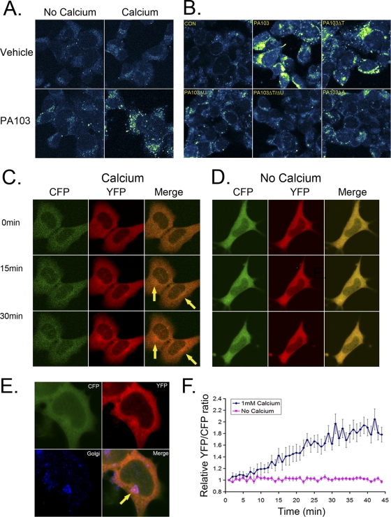
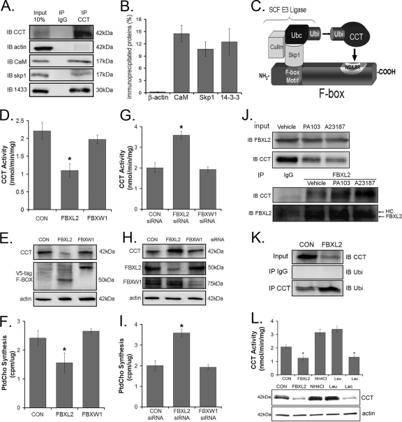
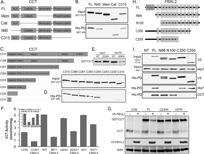
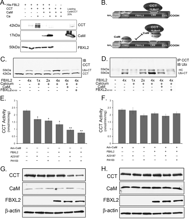
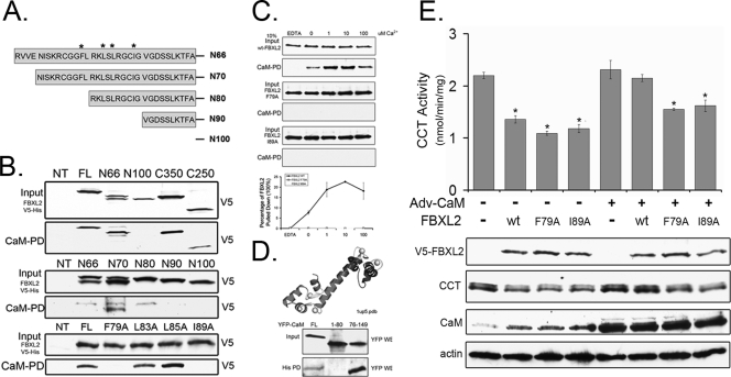
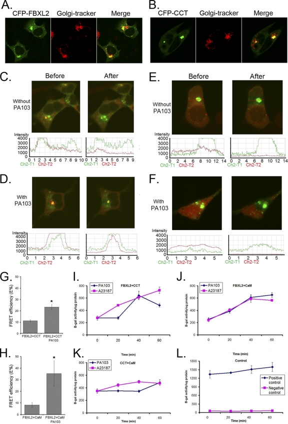
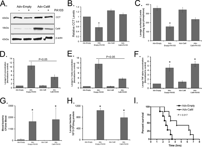
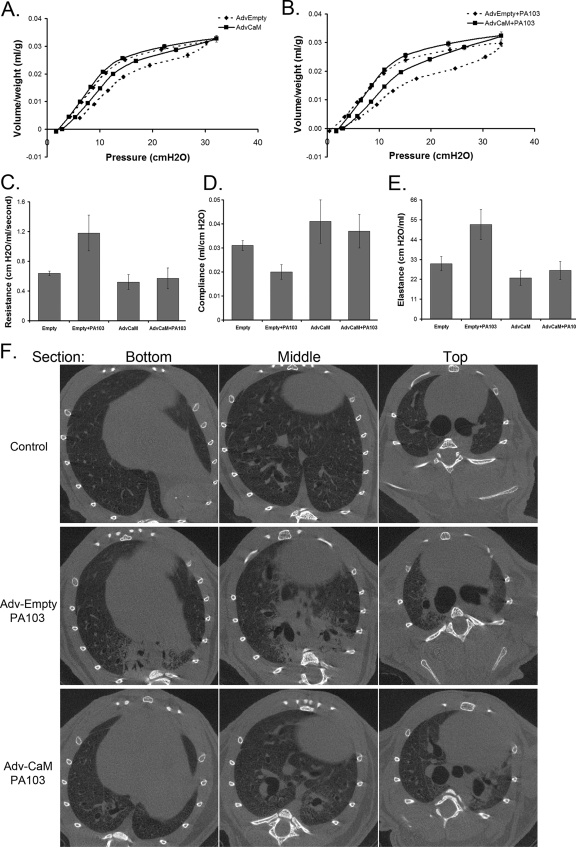
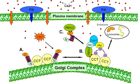
Calmodulin is a universal calcium-sensing protein that has pleiotropic effects. Here we show that calmodulin inhibits a new SCF (Skp1-Cullin-F-box) E3 ligase component, FBXL2. During Pseudomonas aeruginosa infection, SCF (FBXL2) targets the key enzyme, CCTα, for its monoubiquitination and degradation, thereby reducing synthesis of the indispensable membrane and surfactant component, phosphatidylcholine. P. aeruginosa triggers calcium influx and calcium-dependent activation of FBXL2 within the Golgi complex, where it engages CCTα. FBXL2 through its C terminus binds to the CCTα IQ motif. FBXL2 knockdown increases CCTα levels and phospholipid synthesis. The molecular interaction of FBXL2 with CCTα is opposed by calmodulin, which traffics to the Golgi complex, binds FBXL2 (residues 80 to 90) via its C terminus, and vies with the ligase for occupancy within the IQ motif. These observations were recapitulated in murine models of P. aeruginosa-induced surfactant deficiency, where calmodulin gene transfer reduced FBXL2 actions by stabilizing CCTα and lessening the severity of inflammatory lung injury. The results provide a unique model of calcium-regulated intermolecular competition between an E3 ligase subunit and an antagonist that is critically relevant to pneumonia and lipid homeostasis.


Figures

Fig. 1.
Fig. 1.
P. aeruginosa PA103 triggers Ca2+ influx in murine lung epithelia. (A and B) MLE cells were cultured with Ca2+ Green dye with or without PA103 (MOI, 10; 1 h) or with 1 mM Ca2+ followed by staining with Ca2+ Green dye (1:2,000) for 30 min; cells were then washed with PBS and fixed with 4% paraformaldehyde. A 488-nm wavelength was used to excite the Ca2+ Green dye, with visualization of fluorescence emission. In panel B, cells were also infected (MOI, 10; 1 h) with or without PA103 mutants defective in expression of either the TTSS gene (ExsA; ΔA), exotoxin U (ΔU), or ExoT (ΔT) or mutants harboring a double deletion for ExoU and ExoT (ΔT/ΔU). Cells were stained with Ca2+ Green dye, washed, and fixed, followed by visualization of fluorescence emission. (C and D) Cells were infected with baculovirus encoding cameleon prior to culture in medium replete with Ca2+ (1 mM) or depleted of Ca2+, followed by PA103 infection (MOI, 10; 1 h) prior to processing for visualization of fluorescent CFP and YFP signals. (E) Following PA103 infection, cells were washed, fixed, and immunostained using anti-Golgi 97 antibody (1:200) to visualize the Golgi complex. (F) Cells infected with baculovirus encoding cameleon and PA103 as described for panel C were processed for analysis of fluorescent CFP and YFP signals.
Fig. 2.
Fig. 2.
Identification of an E3 ligase for CCTα. (A and B) CCTα was immunoprecipitated using 2 μg of CCTα antibody from 100 μg of MLE cell lysates and processed for CCT, β-actin, CaM, Skp1, and 14-3-3 immunoblotting (A), and bands were quantitated (B). (C) Cartoon illustrating a classic model of the SCF-E3 ligase complex substrate engagement. (D to F) Two micrograms of each plasmid encoding two F-box proteins was transfected into cells (1 × 10⁶) by nucleofection. Twenty-four hours later, cells were lysed and assayed for CCT activity (D), immunoblotting (E), or phosphatidylcholine synthesis (F). (G to I) Cells (1 × 10⁶) were transfected with 0.2 nmol of scrambled (CON) RNA, FBXL2 siRNA, or FBXW1 siRNA by nucleofection. Forty-eight hours later, cells were lysed and processed for CCT activity (G), immunoblotting (H), and phosphatidylcholine synthesis (I). (J) Cells were treated either with vehicle, PA103 (MOI, 10; 1 h), or A23187 (10 nM) for 4 h. Cells were lysed, and 10 μg of lysate was resolved by SDS-PAGE prior to FBXL2 or CCTα immunoblotting (upper panel). FBXL2 was immunoprecipitated in 200 μg of lysate and processed for CCTα or FBXL2 immunoblotting (lower panel). (K) Cells were transfected with either empty vector or FBXL2 plasmid and lysed; 10 μg of lysate was resolved by SDS-PAGE prior to CCTα immunoblotting (upper panel). CCTα was immunoprecipitated in 200 μg of lysate and processed for ubiquitin immunoblotting (lowest panel). (L) Cells (1 × 10⁶) were transfected with 2 μg of FBXL2 and treated with vehicle, the lysosomal inhibitors leupeptin (1:1,000 dilution) or NH4Cl (10 mM), or the proteasomal inhibitor lactacystin (1:1,000 dilution). Twenty-four hours later, cells were lysed, and assays of CCT activity (upper) and CCTα mass (lower) were performed.
Fig. 3.
Fig. 3.
FBXL2 interaction with CCTα. (A) Map of GST-CCTα mutants. Dashed lines represent deleted motifs. (B) Five micrograms of plasmids encoding GST-tagged mutants (mapped in panel A) were expressed in MLE cells (3 × 10⁶), and 24 h later cells were lysed and processed for GST immunoblotting (upper panel) or purification on His-V5-tagged FBXL2 complexed on cobalt affinity beads prior to GST immunoblotting (lower panel). (C) Map of CCTα mutants lacking motifs within its membrane binding domain. (D) The mutants in panel C were processed similar to those shown in panel B for GST immunoblotting of lysates (upper panel) or after affinity purification (lower panel). His-PD, His-tagged pulldown product. (E) Five micrograms of plasmids encoding either GST-CCT WT or GST-CCTQ243A was expressed in cells (3 × 10⁶), and 24 h later cells were lysed and processed for GST immunoblotting after purification with His-V5-tagged FBXL2 cobalt affinity beads. (F and G) Cells were cotransfected with 3 μg of plasmids encoding FBXL2 and 2 μg of plasmids encoding either WT GST-CCT, GST-CCTQ243A, or GST-CCTK57R and then processed for CCT activity (F) or immunoblotting (G). The inset in panel F shows CCT activity as a percentage of control after FBXL2 expression. (H) Map of FBXL2 mutants. (I) His-V5-FBXL2 mutants were expressed in cells, and lysates were processed for V5 and Skp1 immunoblotting or purified on cobalt affinity beads (His-PD) prior to immunoblotting for V5, Skp1, and CCTα.
Fig. 4.
Fig. 4.
SCF (FBXL2) ubiquitination of CCTα is Ca2+ dependent and opposed by CaM. (A) FBXL2-agarose beads (used as bait) were generated and incubated with combinations of 1 μg purified CCTα or CaM with or without Ca2+. After washing of beads (150 mM NaCl, 0.1% Triton X-100), proteins were eluted and resolved by SDS-PAGE followed by CCT, CaM, and V5 immunoblotting. (B) Cartoon illustrating the proposed FBXL2 targeting mechanism. (C) In vitro ubiquitination of CCTα and immunoblotting. Purified CCTα was incubated with the full complement of immunoprecipitated Cullin 1, Skp1, Rbx1, and FBXL2 from cells, plus ubiquitin (∼8.5 kDa) and ATP. Reactions proceeded in the presence or absence of excess Ca2+, CaM, or the FBXL2N100 mutant. Reaction products were processed for CCTα immunoblotting. (D) Coimmunoprecipitation of monoubiquitinated CCTα. CCTα was immunoprecipitated from the in vitro ubiquitination reactions shown in panel C and processed for ubiquitin immunoblotting. (E to H) Cells (4 × 10⁶) were infected with Adv-empty or Adv-CaM (MOI, 40) for 12 h prior to harvest and transfection with FBXL2 plasmid (5 μg) for an additional 24 h. Cells were then infected with PA103 (MOI, 10) for 1 h or treated with A23187 (10 nM) for 4 h prior to analysis of CCT activity (E and F) or immunoblotting (G and H).
Fig. 5.
Fig. 5.
FBXL2 interaction with CaM. (A) Map of FBXL2 mutants. (B) The FBXL2 truncation mutants shown in Fig. 3H, shown in panel A here, or FBXL2 point mutants were expressed in MLE cells, and lysates were processed for V5 immunoblotting (top panel, each pair) or after incubation and elution with CaM-Sepharose beads (40 μl; lower panel, each pair). (C) Cells (4 × 10⁶) were transfected with 5 μg of plasmids encoding WT-V5-FBXL2, V5-FBXL2-F79A, or V5-FBXL2-I89A. Twenty-four hours later, cells were harvested and 100 μg of cell lysate was incubated with CaM-Sepharose beads at different Ca2+concentrations. After extensive rinsing, elusion products were resolved by SDS-PAGE prior to V5 immunoblotting. Levels of V5 proteins were quantified using densitometry and graphed (lowest panel). Data represent two independent experiments. (D) Crystal structure of CaM, showing the NH2 domain (residues 1 to 80) and the carboxyl-terminal domain (residues 76 to 149). Four Ca2+ ions are bound to two homologous domains composed of two helix-loop-helix motifs, connected by a 5-residue flexible linker (black). YFP-tagged full-length (FL) CaM, CaM containing the first 80 residues (1 to 80), or a stretch of carboxyl-terminal 73 residues (76 to 149) was coexpressed in cells with His-V5-tagged FBXL2. Lysates from transfectants were resolved by SDS-PAGE prior to YFP immunoblotting (upper panel), or lysates were purified on Talon cobalt affinity beads prior to processing for YFP immunoblotting (lower panel). (E) Cells (4 × 10⁶) were plated in 100-mm dishes for 24 h, infected with Ad-CaM or an empty vector (Ad-Con) at an MOI of 40 for 12 h, and cells were then harvested and transfected with 5 μg of plasmid encoding WT-FBXL2, FBXL2-F79A, or FBXL2-I89A for an additional 24 h. Cells were then assayed for CCT activity (upper panel) or immunoblotting (lower panel).
Fig. 6.
Fig. 6.
P. aeruginosa degradation of CCTα involves molecular interactions of FBXL2 and CaM within the Golgi complex. (A and B) MLE cells (2 × 10⁵) were transfected with CFP-FBXL2 or CFP-CCTα plasmids (1 μg) and immunostained with anti-Golgi complex (anti-Golgi 97) antibodies. (C to F) Cells were cotransfected with CFP-FBXL2/YFP-CCTα (C and D) or CFP-FBXL2/YFP-CaM (E and F) for 24 h. Cells were then infected with or without PA103 at an MO1 of 10 for 1 h. Cells were washed with PBS and fixed with 4% paraformaldehyde for 20 min and observed using a confocal microscope. The FBXL2-CCT or FBXL2-CaM interaction at the single-cell level was imaged using laser scanning microscopy before and after photobleaching. Shown in the upper sets of panels are single-cell images before and after acceptor photobleaching fluorescence with intensities of YFP and CFP (C to F). (Bottom) The same FRET in each panel was confirmed quantitatively and is shown graphically. FRET efficiencies (E%) were calculated and are graphed (G and H). (I to L) Mammalian two-hybrid assay. Cells (1 × 10⁶) were cotransfected using electroporation with combinations of CCTα-Gal4BD, CaM-Gal4AD, FBXL2-Gal4BD, and FBXL2-Gal4AD plasmids as fusion proteins with a pFR-β-galactosidase reporter vector prior to assays for β-galactosidase activities.
Fig. 7.
Fig. 7.
Adenoviral CaM gene transfer lessens the severity of P. aeruginosa-induced lung inflammation and injury. C57BL/6J mice were administered i.t. with Adv-empty or Adv-CaM (109 PFU/mouse) for 48 h, and 4 mice/group were inoculated with PA103 (1 × 10⁷ CFU/mouse). Mice were euthanized after 1 h. Lungs were lavaged with saline, harvested, and then homogenized; blood was also withdrawn using cardiac puncture. Lung CCTα, CaM, and β-actin were assayed by immunoblotting (A), and the results were analyzed and quantified using ImageJ software (B). (C) Lavage surfactant lipids were extracted and resolved using thin-layer chromatography, and surfactant phosphatidylcholine mass was then assayed. (D to F) An aliquot of lavage samples was centrifuged, and 0.5% fatty acid-free BSA plus EDTA-free protease inhibitor cocktail was added to the supernatant. Forty microliters of supernatant was assayed for cytokines by using antibodies to IL-1β, IL-6, and tumor necrosis factor (TNF) alpha for immunoblotting, and the results were analyzed and quantified using ImageJ software. (G and H) Blood and whole-lavage diluents were plated on TSB agar plates to determine CFU. (I) Replication-deficient Ad5 alone or Adv-CaM (109 PFU/mouse) was instilled i.t. on day 1, after which animals were allowed to recover for 48 h. Following recovery, mice were deeply anesthetized, and 7 mice/group were inoculated with 1 × 10⁷ CFU PA103. Mice were carefully monitored over time; moribund, preterminal animals were immediately euthanized and recorded as deceased. Kaplan-Meier survival curves were generated using Prism software. *, P < 0.05 versus control.
Fig. 8.
Fig. 8.
Adenoviral CaM gene transfer improves lung mechanics after P. aeruginosa infection. (A and B) Replication-deficient adenovirus (AdvEmpty) alone or Adv-CaM (109 PFU/mouse) was instilled i.t. on day 1, after which animals were allowed to recover for 48 h. Following recovery, mice were deeply anesthetized, followed by infection with P. aeruginosa PA103 (107 CFU/mouse, i.t.) for 1 h. Animals were then mechanically ventilated, and pressure-volume loops were measured (B). (C to E) Lung resistance, compliance, and elastance (lung stiffness). Each group contained five to six mice. (F to H) In separate studies, mice were given adenovirus and PA103 as described above and processed for micro-CT scanning to visualize lung infiltrates.
Fig. 9.
Fig. 9.
Molecular interplay between FBXL2, its substrate CCTα, and CaM controls phosphatidylcholine (PC) synthesis. (A) CCTα is a dimeric lipogenic enzyme normally bound and stabilized by CaM that is activated by membrane binding (e.g., to the Golgi complex). (B) P. aeruginosa increases intracellular Ca2+ (red dots) that recruits FBXL2 to membrane-bound CCTα (lower right); the activated SCF E3 ligase complex (Skp1-Cullin 1-FBXL2) binds and ubiquitinates CCTα, the rate-limiting enzyme required for phosphatidylcholine synthesis. The monoubiquitinated CCTα is targeted for disposal via the endosome-lysosome pathway. (C) Excess CaM competitively rescues CCTα from ubiquitination by directly binding to the FBXL2 E3 ligase via its carboxyl-terminal domain.

Similar articles

Cited by

References

    1. Brandt N. R., Caswell A. H., Brandt T., Brew K., Mellgren R. L. 1992. Mapping of the calpain proteolysis products of the junctional foot protein of the skeletal muscle triad junction. J. Membr. Biol. 127:35–47 - PubMed
    1. Cenciarelli C., et al. 1999. Identification of a family of human F-box proteins. Curr. Biol. 9:1177–1179 - PubMed
    1. Chen B. B., Mallampalli R. K. 2007. Calmodulin binds and stabilizes the regulatory enzyme, CTP: phosphocholine cytidylyltransferase. J. Biol. Chem. 282:33494–33506 - PubMed
    1. Chen B. B., Mallampalli R. K. 2010. Masking of a nuclear signal motif by monoubiquitination leads to mislocalization and degradation of the regulatory enzyme cytidylyltransferase. Mol. Cell. Biol. 29:3062–3075 - PMC - PubMed
    1. Costelli P., et al. 2005. Ca2+-dependent proteolysis in muscle wasting. Int. J. Biochem. Cell Biol. 37:2134–2146 - PubMed

Publication types

MeSH terms